. 2011 Jun 29;31(26):9772-86.
doi: 10.1523/JNEUROSCI.1604-11.2011.

The master negative regulator REST/NRSF controls adult neurogenesis by restraining the neurogenic program in quiescent stem cells

Affiliations

The master negative regulator REST/NRSF controls adult neurogenesis by restraining the neurogenic program in quiescent stem cells

Zhengliang Gao et al. J Neurosci. .

Abstract

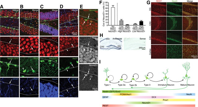
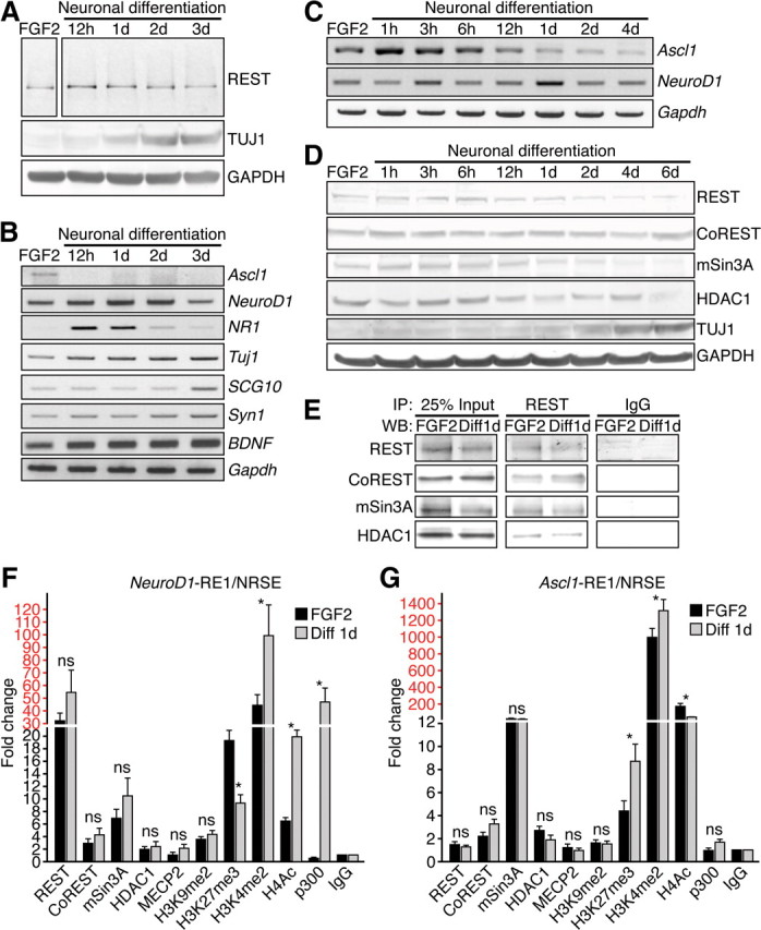
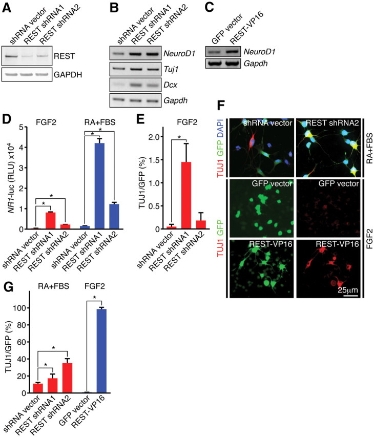
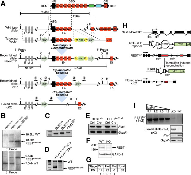
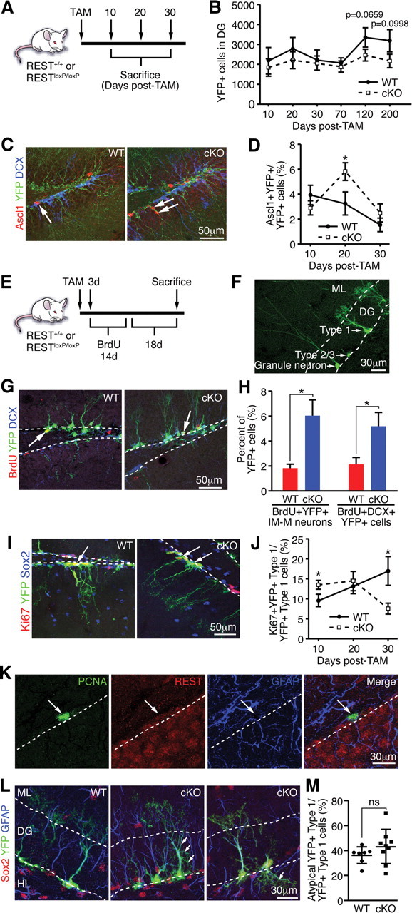
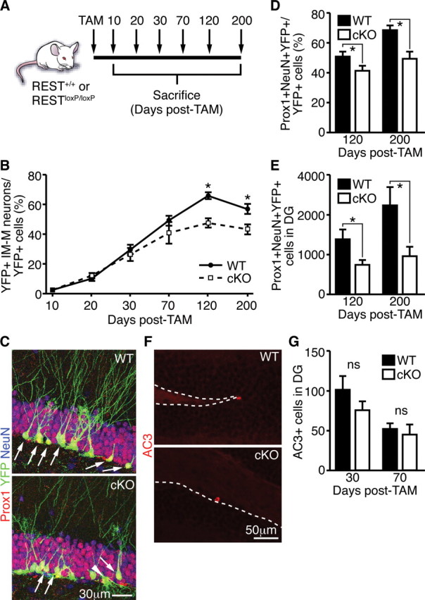
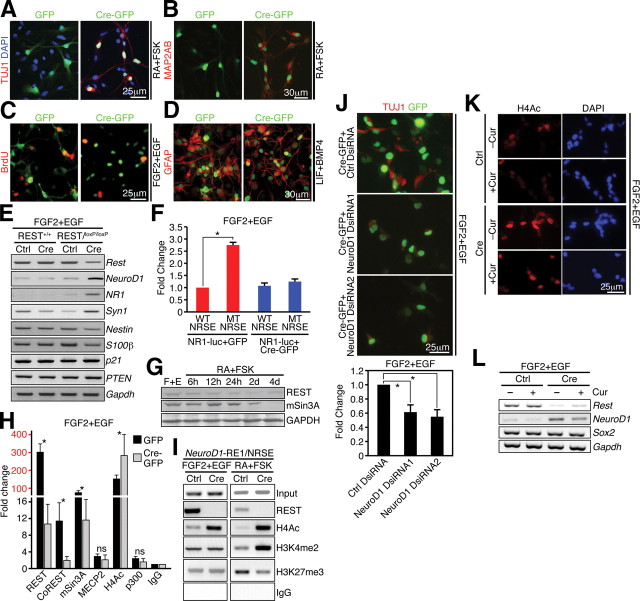
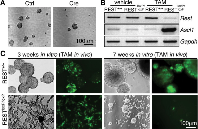
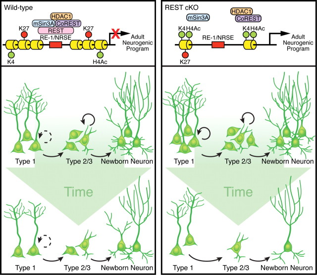
Transcriptional regulation is a critical mechanism in the birth, specification, and differentiation of granule neurons in the adult hippocampus. One of the first negative-acting transcriptional regulators implicated in vertebrate development is repressor element 1-silencing transcription/neuron-restrictive silencer factor (REST/NRSF)--thought to regulate hundreds of neuron-specific genes--yet its function in the adult brain remains elusive. Here we report that REST/NRSF is required to maintain the adult neural stem cell (NSC) pool and orchestrate stage-specific differentiation. REST/NRSF recruits CoREST and mSin3A corepressors to stem cell chromatin for the regulation of pro-neuronal target genes to prevent precocious neuronal differentiation in cultured adult NSCs. Moreover, mice lacking REST/NRSF specifically in NSCs display a transient increase in adult neurogenesis that leads to a loss in the neurogenic capacity of NSCs and eventually diminished granule neurons. Our work identifies REST/NRSF as a master negative regulator of adult NSC differentiation and offers a potential molecular target for neuroregenerative approaches.


Figures

Figure 1.
Figure 1.
REST has biphasic expression in adult hippocampal NSCs and mature granule neurons. A, B, Immunostaining of REST, Nestin–GFP, and Sox2 (markers of quiescent NSCs and transit-amplifying progenitors) and GFAP (a marker of quiescent NSCs). Insets show colocalization of REST with these markers: REST+/Nestin--GFP+/GFAP+ and REST+/Sox2+/GFAP+ type 1 NSCs (A, B, arrows); REST+/Sox2+/GFAP type 2a transit-amplifying progenitor cell (B, arrowhead). C, Immunostaining of REST, NeuroD1, and NeuN (a marker of mature neurons). D, Immunostaining of REST, Ascl1–GFP, and GFAP. REST is expressed in a subset of Ascl1–GFP+ cells. Inset shows REST+/Ascl1–GFP+/GFAP+ cells (arrow) and REST+/Ascl1–GFP+/GFAP cells (arrowhead). E, REST is highly expressed in mature granule neurons and some low NeuroD1+ cells (arrowhead), whereas it is only expressed in a minor subset of high NeuroD1+ cells (arrows). Dotted lines indicate border between SGZ and hilus. F, Assessment of REST and NeuroD1 colocalization demonstrated an inverse relationship between the expression of REST and NeuroD1, implying a repressive role for REST in NeuroD1 expression during adult neurogenesis. Error bars represent ± SEM. G, H, Immunostaining (G) and RNA in situ hybridization (H) of 6-week-old adult mouse brain showing that REST is broadly expressed in postmitotic neurons (NeuN+) in DG, CA3, CA1, and cortex. I, Summary of REST expression with different markers during adult hippocampal neurogenesis.
Figure 2.
Figure 2.
The REST complex represses neuronal genes in HCN cells. A–C, Western blot analysis (A) or RT-PCR analysis (B, C) of REST and/or its target genes after RA-induced (1 nm) and FSK-induced (5 nm) neuronal differentiation. Gapdh was used as a normalization control. D, Western blot analysis of REST and its corepressors CoREST, mSin3A, and HDAC1 during neuronal differentiation. TUJ1 was used as a positive control for neuronal differentiation, and GAPDH was used as a loading control. E, Coimmunoprecipitation experiments showing the interaction of REST with its corepressors CoREST, mSin3A, and HDAC1. WB, Western blot. F, G, The occupancy of REST, cofactors, and histone modifications at the NeuroD1– and Ascl1–RE1/NRSE sites in HCN cells under proliferation conditions (FGF2) and under differentiation conditions (RA–FSK for 24 h). Relative enrichment of factor binding was calculated as fold changes relative to IgG control (n = 3) under the same conditions after normalizing to the respective input control. Different scales are demarked by different colors (black and red). Error bars represent ± SEM. *p ≤ 0.05 by two-tailed Student's t test. NS, Not significant.
Figure 3.
Figure 3.
Knockdown of REST results in de-repression of pro-neuronal genes and accelerates the neuronal phenotype. A, Confirmation of REST shRNA knockdown in HCN cells. Two different REST shRNAs resulted in efficient knockdown of endogenous REST protein. GAPDH was used as a normalization control. B, De-repression of neuronal target genes during REST shRNA knockdown in differentiation conditions (RA–FSK for 24 h). C, Upregulation of NeuroD1 during expression of REST–VP16 under proliferation conditions (FGF2). D, Luciferase reporter assays showing REST shRNA-mediated de-repression of promoter reporter activity of NR1 (a REST target gene) in both proliferating (FGF2) and differentiation (RA–FBS for 24 h) conditions. E, Quantification of REST shRNA-mediated neuronal differentiation (percentage TUJ1) in proliferation conditions (FGF2). F, shRNA knockdown of REST enhanced neuronal differentiation when HCN cells were primed with RA and FBS for 24 h, and introduction of REST–VP16 resulted in precocious differentiation under proliferating conditions (FGF2). G, Quantification of REST shRNA- or REST–VP16-mediated neuronal differentiation (percentage TUJ1) in differentiation conditions (RA–FBS) and proliferation conditions (FGF2), respectively. Error bars represent ± SEM. *p ≤ 0.05 by two-tailed Student's t test.
Figure 4.
Figure 4.
Generation of REST cKO mice. A, Strategy to generate a REST cKO allele. Protein, corresponding exonic structure, targeting vector, and targeted allele are depicted. The FRT site flanked neomycin resistance cassette was removed by crossing to transgenic animals expressing hACTB:FLPe in the germ line. H, HpaI; X, XbaI; NR, N-terminal repressor domain; CR, C-terminal repressor domain; DBD, DNA binding domain. B, Southern blot analysis of WT, RESTneo-loxP/+, and RESTneo-loxP/neo-loxP. Tail DNA was digested with XbaI (5′ probe) and HpaI (3′ probe), and the corresponding WT (16.5/7.5 kb) and targeted (10.5/9.5 kb) bands are indicated for the 5′/3′ probes. C, Genotyping of RESTloxP mice by genomic PCR. The primer set (3 + 4) flanks the 3′ loxP site, resulting in one ∼550 bp product for the WT allele and one ∼650 bp band for the targeted allele. D, Successful recombination with global deletion by CAG–Cre. The primer set (1 + 4) flanks both the 5′ and 3′ loxP sites, resulting in one ∼2.8 kb product for the WT allele and ∼1.1 kb band for the targeted allele. E, F, RT-PCR and Western blot analyses to confirm successful deletion of the REST cKO allele by Ad–Cre-mediated recombination in vitro in RESTloxP/loxP mouse embryonic fibroblast cells (MEFs) and NSCs (data not shown). G, REST+/+ or REST+/loxP; CAG–Cre mice segregate according to Mendelian ratios, whereas RESTloxP/loxP; CAG–Cre mutant is embryonic lethal, recapitulating the previously reported global KO phenotype (Chen et al., 1998). H, Strategy to generate an NSC-specific deletion of REST using an inducible form of Nestin–CreERT2. I, Assessment of TAM-induced recombination frequency in vivo by establishing adult NSC cultures from TAM-treated mice. An NSC line of >95% YFP+ cells was used to assess REST recombination frequency within the YFP+ population during TAM treatment using REST genomic PCR (top 2 panels) and RT-PCR (bottom 2 panels). DNA from non-TAM-treated REST/NRSFloxP/loxP NSCs and DNA from REST/NRSF+/+ NSCs were mixed at ratios (loxP:+) of 9:1, 1:2, 1:3, and 1:9, and PCR was performed with primer set (1 + 2) to produce an amplification standard. DNA from TAM-treated REST/NRSFloxP/loxP NSCs (cKO lane) showed barely detectable amplification (>90% recombination efficiency), and DNA from REST/NRSF+/+ NSCs (WT lane) showed absence of product with primer set (1 + 2) under identical PCR conditions. We confirmed positive amplification of the floxed allele cKO with primer set (1 + 4) in DNA from TAM-treated REST/NRSFloxP/loxP NSCs but not from REST/NRSF+/+ NSCs.
Figure 5.
Figure 5.
Deletion of REST in vivo leads to precocious neuronal differentiation and impaired maintenance of type 1 cells. A, Schematic of TAM treatment and harvesting of brain tissue. B, No significant change in the number of total YFP+ cells was observed at 10 d (p = 0.3618), 20 d (p = 0.4252), 30 d (p = 0.7903), and 70 d (p = 0.6281) after TAM, but there was a strong decreasing trend in REST cKO compared with WT at 120 d (p = 0.0659) and 200 d (p = 0.0998) after TAM. C, Representative images of an Ascl1+/YFP+ cell in WT (arrow, left) and Ascl1+/YFP+ cells in REST cKO (arrows, right). DCX (blue) is a marker for immature granule neurons. D, REST ablation transiently increases the percentage of Ascl1+/YFP+ cells at 20 d, but not at 10 or 30 d, after TAM. E, Schematic of TAM and BrdU treatment. F, Representative morphology of YFP+ type 1, 2/3 cells, and granule neurons in the DG. ML, Molecular layer. G, Representative images of a BrdU+/YFP+ cell in WT (arrow, left) and a BrdU+/DCX+/YFP+ cell in REST cKO (arrow, right). H, REST ablation accelerates the progression NSCs to immature and mature (IM-M) neurons as shown by morphological phenotyping of BrdU+/YFP+ cells and increased percentage of BrdU+/DCX+/YFP+ cells of the total YFP+ cells in REST cKO mice compared with WT. I, Representative images of a Ki67+/YFP+ type 1 cell in WT (arrow, left) and Ki67+/YFP+ cells in REST cKO (arrows, right). Sox2 (blue) stains type 1 and 2 cells. J, REST ablation in adult NSCs transiently increases type 1 cell proliferation at 10 d after TAM, determined by quantifying Ki67+/YFP+ type 1 cells, which are quickly depleted, ultimately resulting in a decrease in type 1 cell proliferation 30 d after TAM. K, Immunostaining of REST and cell-type-specific markers showing that REST is expressed in PCNA+/GFAP+ proliferating type 1 cells (arrow). Dotted lines indicate border of SGZ and hilus. L, REST cKO type 1 cells display atypical morphological characteristics (see Results). Representative images of YFP+ type 1 cells in WT and REST cKO mice are shown. Arrows indicate examples of atypical type 1 cell morphology in REST cKO. We confirmed that GFAP (blue) and Sox2 (red) are expressed in YFP+ type 1 cells in both WT and REST cKO. Dotted lines indicate the boundaries of the GCL. ML, Molecular layer; HL, hilus. M, Quantification of the percentage of atypical YFP+ type 1 cells in WT (n = 7) compared with REST cKO (n = 9). Error bars represent ±SEM. *p ≤ 0.05 by two-tailed Student's t test. If not indicated by * or ns, then not significant.
Figure 6.
Figure 6.
Decreased granule neuron production in REST cKO mice over time. A, Schematic of TAM treatment and harvesting of brain tissue. B, Ablation of REST in adult NSCs resulted in decreased production of immature and mature granule neurons (IM-M) at 120 and 200 d after TAM. There was no significant change between REST cKO and WT at 10, 20, 30, and 70 d after TAM. C, Representative images of Prox1+/NeuN+/YFP+ cells in the SGZ of WT (arrows, top) or REST cKO (arrows, bottom). Note an YFP+ type 1 cell with atypical morphology in REST cKO (arrowhead). D, E, In contrast to the earlier transient increase in immature and mature neurons (Fig. 5H) and consistent with a functional depletion of the stem cell pool, REST ablation in adult NSCs resulted in decreased production of Prox1+/NeuN+/YFP+ mature granule neurons at 120 and 200 d after TAM. F, Representative images of AC3+ cells in WT and REST cKO mice. Dotted line indicates the border between SGZ and hilus. G, No change in AC3+ cells was observed between WT and REST cKO mice. Error bars represent ±SEM. *p ≤ 0.05 by two-tailed Student's t test. If not indicated by * or ns, then not significant.
Figure 7.
Figure 7.
REST is required in adult NSCs to control stage-specific neuronal gene expression in primary NSCs. A, B, Ad–Cre-mediated ablation of REST in SVZ and hippocampal NSCs promotes precocious neuronal differentiation when primed with RA–FSK as indicated by morphology and neuronal marker staining (TUJ1+ and MAP2AB+). C, Neural progenitor cell proliferation was decreased in REST cKO after Ad–Cre-mediated ablation in proliferating conditions (FGF2+EGF) compared with either Ad–GFP-infected cKO cells or Ad–Cre-infected WT cells. D, Deletion of REST in astrocyte differentiation media (LIF–BMP-4 for 4 d) results in morphological changes but does not significantly compromise the number of GFAP+ astrocytes. E, During REST deletion, many of its target neuronal genes (NeuroD1, NR1, and Syn1) are upregulated, whereas some NSC (Nestin), glial (S100β), or cell cycle genes (p21 and PTEN) are downregulated or remain unchanged. Gapdh was used as an internal control. F, Luciferase reporter assays showing RE1/NRSE-dependent regulation of NR1 expression, a known REST target. G, REST and mSin3A levels remained unchanged immediately after neuronal induction with RA–FSK but declined over time when compared with cells grown in proliferation conditions (FGF2–EGF). H, ChIP–QPCR analysis showing that REST ablation leads to changes in corepressor binding at the NeuroD1–RE1/NRSE. The occupancy of respective factors was calculated as fold changes relative to IgG control (n = 3) in the same conditions (Ad–GFP or Ad–Cre–GFP infected) after normalizing to the respective input control. Different scales are demarked by different colors (black and red). I, ChIP–PCR of REST and various histone modifications in WT and cKO NSCs. J, NeuroD1 knockdown partially attenuates the precocious differentiation phenotype resulting from REST ablation. For quantification, the level of differentiation in the control (Ctrl) DsiRNA group was set as 1, and the experimental DsiRNA groups (NeuroD1 DsiRNA1 and NeuroD1 DsiRNA2) were then normalized accordingly. K, L, Treatment with curcumin (p300/CBP-specific HAT inhibitor) blocks histone acetylation as indicated by H4Ac immunostaining (K) and upregulation of NeuroD1 after REST ablation (L). Error bars represent ±SEM. *p ≤ 0.05 by two-tailed Student's t test.
Figure 8.
Figure 8.
REST is required for self-renewal of primary mouse NSCs in vitro. A, Ad–Cre-mediated REST ablation in primary mouse NSCs in vitro diminished their colony formation capacity. B, Confirmation of successful ablation of REST in mouse NSCs with TAM treatment in vivo accompanied by a dramatic upregulation of Ascl1 mRNA. C, TAM-induced REST deletion in NSCs in vivo abolished their ability to give rise to sustained neurosphere cultures in vitro. YFP+ NSCs from TAM-treated REST mice failed to form spheres and were depleted with passage over time. Shown in the figure are representative pictures from NSC cultures 3 weeks in vitro (left panels) and 7 weeks in vitro (right panels).
Figure 9.
Figure 9.
Model of REST function during adult neurogenesis. Left, In wild-type mice, REST and its corepressor complexes containing mSin3A, CoREST, and HDAC1 restrict the adult neurogenic program (e.g., Ascl1, NeuroD1, NR1, and other neuronal genes). REST functions to restrain the neurogenic program in quiescent and proliferating NSCs and regulate stage-specific neuronal gene expression, thus ensuring continued neurogenesis throughout life. Right, Loss of REST mediates changes in histone acetylation (H4Ac) and bivalent chromatin (H3K4me2 and H3K9me3) that lead to the activation/dysregulation of neuronal gene expression in NSCs. Consequently, adult NSCs prematurely exit quiescence and precociously differentiate into granule neurons. Over time, the neurogenic capacity of adult NSCs becomes exhausted, ultimately leading to decreased neurogenesis.

References

    1. Ables JL, Decarolis NA, Johnson MA, Rivera PD, Gao Z, Cooper DC, Radtke F, Hsieh J, Eisch AJ. Notch1 is required for maintenance of the reservoir of adult hippocampal stem cells. J Neurosci. 2010;30:10484–10492. - PMC - PubMed
    1. Abrajano JJ, Qureshi IA, Gokhan S, Zheng D, Bergman A, Mehler MF. REST and CoREST modulate neuronal subtype specification, maturation and maintenance. PLoS One. 2009;4:e7936. - PMC - PubMed
    1. Balasubramanyam K, Varier RA, Altaf M, Swaminathan V, Siddappa NB, Ranga U, Kundu TK. Curcumin, a novel p300/CREB-binding protein-specific inhibitor of acetyltransferase, represses the acetylation of histone/nonhistone proteins and histone acetyltransferase-dependent chromatin transcription. J Biol Chem. 2004;279:51163–51171. - PubMed
    1. Ballas N, Battaglioli E, Atouf F, Andres ME, Chenoweth J, Anderson ME, Burger C, Moniwa M, Davie JR, Bowers WJ, Federoff HJ, Rose DW, Rosenfeld MG, Brehm P, Mandel G. Regulation of neuronal traits by a novel transcriptional complex. Neuron. 2001;31:353–365. - PubMed
    1. Ballas N, Grunseich C, Lu DD, Speh JC, Mandel G. REST and its corepressors mediate plasticity of neuronal gene chromatin throughout neurogenesis. Cell. 2005;121:645–657. - PubMed

Publication types

Substances

LinkOut - more resources