. 2012 Mar;19(3):338-46.
doi: 10.1038/gt.2011.96. Epub 2011 Jun 30.

Transcription factor plasmid binding modulates microtubule interactions and intracellular trafficking during gene transfer

Affiliations

Transcription factor plasmid binding modulates microtubule interactions and intracellular trafficking during gene transfer

M A Badding et al. Gene Ther. 2012 Mar.

Erratum in

  • Gene Ther. 2012 Mar;19(3):354

Abstract

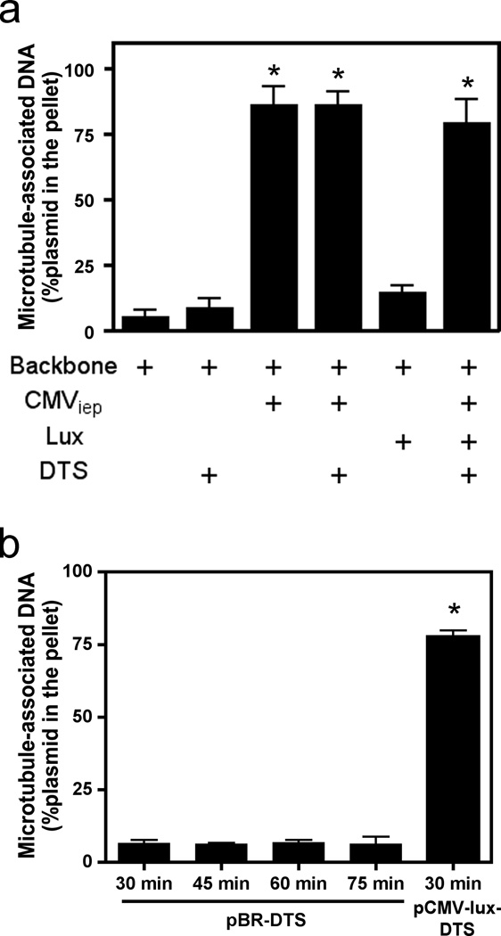
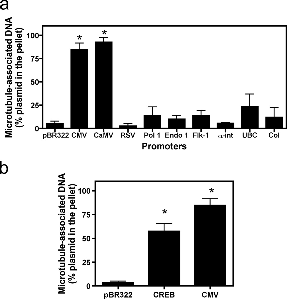
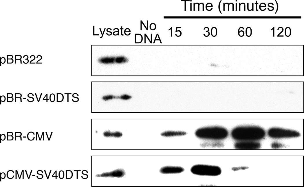
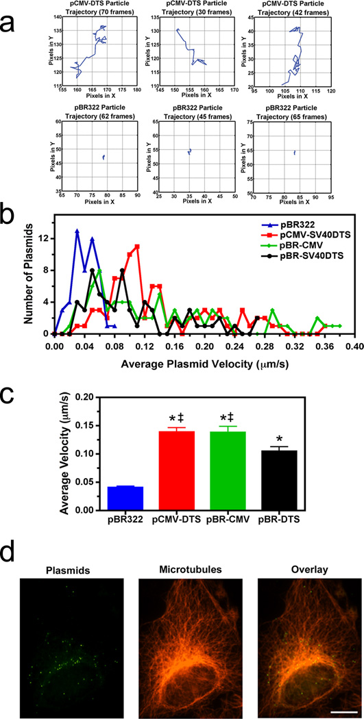
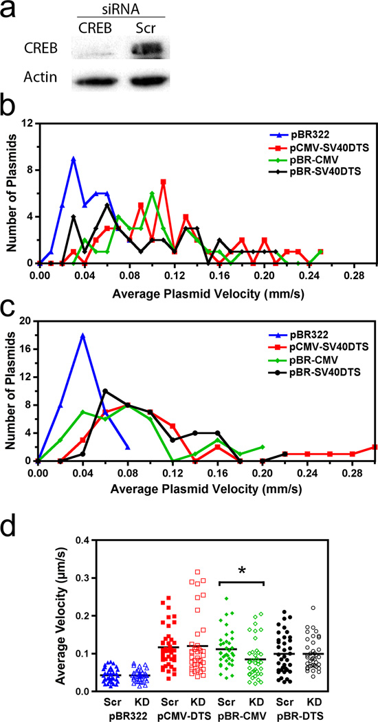
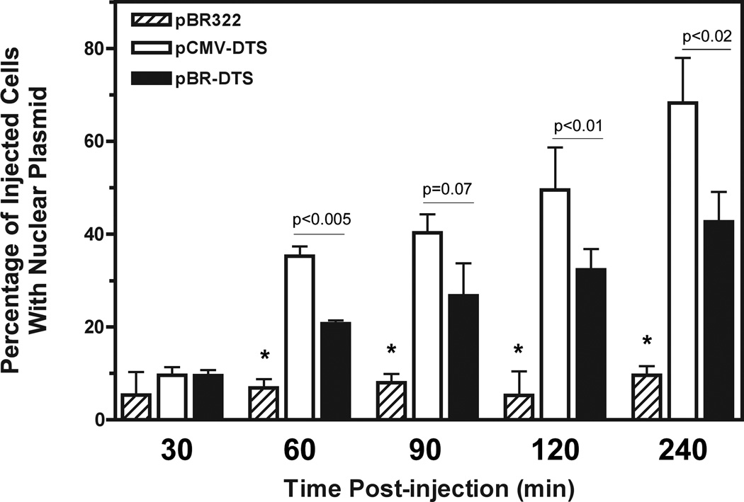
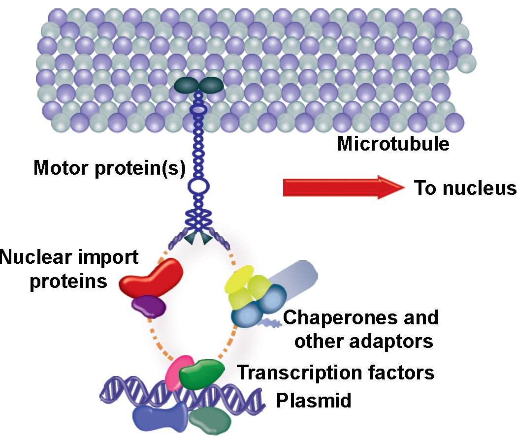
For non-viral gene delivery to be successful, plasmids must move through the cytoplasm to the nucleus in order to be transcribed. While the cytoskeletal meshwork acts as a barrier to plasmid DNA movement in the cytoplasm, the microtubule network is required for directed plasmid trafficking to the nucleus. We have shown previously that plasmid-microtubule interactions require cytoplasmic adapter proteins such as molecular motors, transcription factors (TFs) and importins. However, not all plasmid sequences support these interactions to allow movement to the nucleus. We now demonstrate that microtubule-DNA interactions can show sequence specificity with promoters containing binding sites for cyclic AMP response-element binding protein (CREB), including the cytomegalovirus immediate early promoter (CMV(iep)). Plasmids containing CREB-binding sites showed stringent interactions in an in vitro microtubule-binding assay. Using microinjection and real-time particle tracking, we show that the inclusion of TF binding sites within plasmids permits cytoplasmic trafficking of plasmids during gene transfer. We found that CREB-binding sites are bound by CREB in the cytoplasm during transfection, and allow for enhanced rates of movement and subsequent nuclear accumulation. Moreover, small interfering RNA knockdown of CREB prevented this enhanced trafficking. Therefore, TF binding sites within plasmids are necessary for interactions with microtubules and enhance movement to the nucleus.


Conflict of interest statement

Conflict of Interest

The authors declare no conflict of interest.

Figures

Figure 1
Figure 1. Microtubule spin-down assays showing in vitro interaction of DNA with microtubules
(a) Quantitative analysis of plasmid elements that associate with microtubules. Plasmids containing different sequence elements (the CMV promoter, the luciferase gene, and/or the DTS) were incubated for 30 minutes with cell extract and taxol-stabilized microtubules and subsequently separated over a glycerol cushion by centrifugation. The plasmid contents of the pellets and supernatants were determined by quantitative PCR, and percentage of DNA in the pellet was determined by comparing DNA content in pelleted fractions versus total DNA in both supernatant and pellet fractions combined. (b) Increased incubation times do not change the ability of the DTS to mediate microtubule interactions. pBR322-DTS was incubated with microtubules in the presence of cell extract for 30, 45, 60, and 75 minutes and subsequently centrifuged and quantified by quantitative PCR as in a. Mean DNA concentrations from three independent experiments, preformed in duplicate, are shown ± st. dev. CMV, Cytomegalovirus; Lux, luciferase gene; DTS, Simian Virus 40 DNA nuclear targeting sequence.
Figure 2
Figure 2. Microtubule binding by plasmids containing various eukaryotic promoters
(a) Not all promoters mediate microtubule-DNA interactions. The following promoters were tested in the microtubule spin-down assay as in Figure 1: pBR322 (backbone plasmid, no promoter), CMViep, the Rous sarcoma virus LTR (RSV), the endothelian I promoter (endo), the VEGF receptor promoter (FLK-1), the alpha integrin promoter (αint), the human ubiquitin C promoter (UbC), the 45S RNA polymerase I promoter (Pol I), the human collagen A promoter (Col), and the 35S promoter of Cauliflower Mosaic virus (CaMV). (b) A single CREB-binding site is sufficient to mediate microtubule-plasmid interactions. Plasmids (pBR322; CREB, a plasmid with just one CREB binding site; or CMV, with just the CMViep) were used in the spin-down assay as in Figure 1a. Mean DNA concentrations from three independent experiments, preformed in duplicate, are shown ± st. dev. *, p < 0.0001.
Figure 3
Figure 3. Binding of CREB by plasmids containing the CMViep during gene transfer
Biotinylated plasmids were electroporated into A549 cells, and at the indicated times post transfection, formaldehyde was added to cross-link the DNA-protein complexes, cells were lysed, complexes were pulled down with streptavidin-coated beads, cross-links were reversed by boiling beads with Laemmli Sample buffer, and the resulting lysates were run in Western blots using antibodies against CREB. “Lysate” represents crude lysates for each sample, with no bead precipitation. The “No DNA” lane contains pulled down lysates from untransfected cells, showing only background CREB detection. Experiments were performed in duplicate and repeated four times.
Figure 4
Figure 4. Requirement of promoter or enhancer sequences for plasmid movement in the cytoplasm
(a) Representative traces for individual plasmid trajectories. Quantum dot-labeled plasmids were cytoplasmically microinjected into adherent A549 cells and imaged at 1-second intervals over 5–10 minutes. The traces of representative negative control (pBR322) and positive control (pCMV-DTS) plasmids in injected cells are shown. Plasmid trajectories were created using the PolyParticleTracker software downloaded for use in MATLAB, and all plot areas are mapped to 25 × 25 pixel areas. (b) Average cytoplasmic velocity of individual microinjected plasmids. Movement of individual DNA particles, shown in a, were tracked for up to 10 minutes using time-lapse imaging (1 frame/second). The average velocity of each was determined using particle tracking software (PolyParticleTracker, MATLAB), and the frequency distribution histograms are plotted as the number of plasmids moving at certain velocities for each construct. At least 50 particles were tracked per construct in 3–5 separate experiments. (c) Individual microinjected plasmid velocities were averaged for each of the four constructs. Error bars represent means + st. dev. ‡, p<0.05 compared to pBR-DTS; *, p<0.001 compared to pBR322. (d) Colocalization of Quantum dot-labeled plasmids with microtubules. Cells were microinjected with biotin-PNA-pCMV-DTS plasmids, fixed 20 minutes post-injection, and labeled with anti-tubulin antibodies and streptavidin-conjugated Quantum dots to label plasmids. Cells were imaged using a 100x objective by deconvolution microscopy and a representative deconvolved Z slice is shown. Scale bar, 10 µm.
Figure 5
Figure 5. Enhanced plasmid intracellular movement is prevented with siRNA against CREB
(a) CREB knockdown via RNA interference. Western blot images show CREB protein levels are reduced around 75% in cell lysates after 48 hours incubation with siRNA against CREB (Ambion, Austin, TX) compared to cells transfected with a negative control scramble RNA. (b) Average cytoplasmic velocity of individual microinjected plasmids in scrambled RNA-transfected cells. Movement of individual DNA particles were tracked for 10 minutes and velocities analyzed as in Figure 4. (c) Average cytoplasmic velocity of individual microinjected plasmids in CREB siRNA-transfected cells. Plasmids were imaged and velocities recorded as in b. (d) Scatter plot showing the range of the average velocities for each of the four constructs. Bars represent median velocity for each construct. *, p<0.001. KD, CREB knockdown cells; SC, scrambled control.
Figure 6
Figure 6. CREB-binding plasmids with a DTS sequence have enhanced nuclear localization at earlier time points than plasmids containing the DTS alone
Negative control plasmid pBR322, pBR-DTS, or pCMV-DTS plasmids labeled with CY3-PNA were injected into A549 cells then incubated for the indicated times. Injected cells were imaged and scored for nuclear import. Experiments were performed in triplicate and at least 50–100 cells were scored per time point. *, p<0.01 compared to pCMV-DTS and pBR-DTS for each time point. Error bars represent means + st. dev.
Figure 7
Figure 7. Possible protein interactions in the plasmid trafficking complex
Our model of plasmid interaction with microtubules is not a direct one, and involves transcription factor binding to unique sequences on the plasmid (i.e., CREB binding to CMViep). One or more of the transcription factor NLSs are bound by importins, which are bound by the motor protein dynein and can move along microtubules toward the nucleus for import. Other adaptor molecules, such as chaperones, MAPs (microtubule-associated proteins), and nuclear import proteins may be part of this larger complex as well. TF, transcription factor; NLS, nuclear localization signal; MT, microtubule.

References

    1. Mesika A, Kiss V, Brumfeld V, Ghosh G, Reich Z. Enhanced intracellular mobility and nuclear accumulation of DNA plasmids associated with a karyophilic protein. Hum Gene Ther. 2005;16:200–208. - PubMed
    1. Vaughan EE, Dean DA. Intracellular trafficking of plasmids during transfection is mediated by microtubules. Mol Ther. 2006;13:422–428. - PMC - PubMed
    1. Leopold PL, Kreitzer G, Miyazawa N, Rempel S, Pfister KK, Rodriguez-Boulan E, et al. Dynein- and microtubule-mediated translocation of adenovirus serotype 5 occurs after endosomal lysis. Hum Gene Ther. 2000;11:151–165. - PubMed
    1. Vaughan EE, DeGiulio JV, Dean DA. Intracellular trafficking of plasmids for gene therapy: mechanisms of cytoplasmic movement and nuclear import. Curr Gene Ther. 2006;6:671–681. - PMC - PubMed
    1. Wilson GL, Dean BS, Wang G, Dean DA. Nuclear import of plasmid DNA in digitonin-permeabilized cells requires both cytoplasmic factors and specific DNA sequences. J Biol Chem. 1999;274:22025–22032. - PMC - PubMed

Publication types

Substances