. 2011 Mar;56(2):180-9.
doi: 10.4103/0019-5154.80413.

Vitiligo: a review of some facts lesser known about depigmentation

Affiliations

Vitiligo: a review of some facts lesser known about depigmentation

James J Nordlund. Indian J Dermatol. 2011 Mar.

Abstract

Vitiligo is a disorder that causes the destruction of melanocytes. It has three important factors underlying this destruction. The depigmented skin has many aberrant functions such as a muted response to contact allergens, a phenomenon also seen in mice that depigment. The white skin of those with vitiligo does not form non-melanoma skin cancers although the white skin of albinos, which has a similar color as vitiligo, is highly susceptible to skin cancer.

Keywords: Vitiligo; albinos; contact allergen; non-melanoma skin cancer.


Conflict of interest statement

Conflict of Interest: Nil.

Figures

Figure 1
Figure 1
Depigmentation of the fingers and dorsum of the hands, classical, early manifestations of vitiligo
Figure 2
Figure 2
Depigmentation of the feet, typical early manifestation of vitiligo
Figure 3
Figure 3
Depigmentation around the eyes (partially blocked), the nares and mouth, all classical and early manifestations of bilateral vitiligo
Figure 4
Figure 4
Extensive depigmentation of a woman which illustrates the symmetrical distribution of classical bilateral vitiligo
Figure 5
Figure 5
Unilateral (segmental) vitiligo affecting one side of the face of a young boy. Note that the depigmentation does not correspond to a dermatome
Figure 6
Figure 6
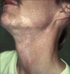
Unilateral depigmentation (segmental vitiligo) affecting the neck of a woman. Note the very similar distribution of depigmentation in the two men pictured in Figures 7 and 8
Figure 7
Figure 7
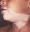
Unilateral depigmentation (segmental vitiligo) of the neck of a man which is similar to the depigmentation seen in Figures 6 and 8
Figure 8
Figure 8
Unilateral depigmentation (segmental vitiligo) on the neck of a man with a distribution similar to that in Figures 6 and 7
Figure 9
Figure 9
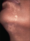
Nevus depigmentosus on the neck of a boy. The pigmentary abnormality was present at birth in the patient
Figure 10
Figure 10
Nevus depigmentosus on the neck of a man. The pigmentary abnormality was present at birth in the patient
Figure 11
Figure 11
Unilateral depigmentation (segmental vitiligo) on the anterior chest and upper part of the right arm in a young woman. The distribution of pigment loss is very similar in this patient and the one given in Figure 12
Figure 12
Figure 12
Unilateral depigmentation (segmental vitiligo) on the anterior chest and upper part of the right arm in a girl. The distribution of pigment loss is very similar in this patient and the one shown in Figure 11
Figure 13
Figure 13
A nevus depigmentosus in a young girl with a distribution of pigment loss similar to the unilateral vitiligo seen in Figures 11 and 12
Figure 14
Figure 14
An Indian woman with veloce vitiligo. She lost almost all of her pigmentation on the skin and hair over a period of some months
Figure 15
Figure 15
The epidermis is composed of three main cell types that work together like a troika
Figure 16
Figure 16
A woman treated with monobenzone, who developed a contact allergy. The dermatitis is exclusively confined to the pigmented skin
Figure 17
Figure 17
A man treated with monobenzone, who developed a contact allergy. The dermatitis is exclusively confined to the pigmented skin
Figure 18
Figure 18
Mice with mivit/mivit gene. The mouse on the left has a piebald band on the leg but a normal pigmented pelage in early life. The mouse on the right is undergoing depigmentation with loss of melanocytes from the epidermis and hair follicles
Figure 19
Figure 19
The neck of an African woman who had vitiligo for over 20 years. She was a farmer and worked in the sun daily. Note the elastosis of the neck, but the normal overlying white epidermis
Figure 20
Figure 20
The hands of the African woman shown in Figure 19, who had vitiligo for over 20 years. She was a farmer and worked in the sun daily
Figure 21
Figure 21
The arms of a woman with piebaldism (c-kit). She has marked sun damage and many actinic keratoses, mostly in the depigmented patches on her arms
Figure 22
Figure 22
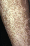
The leg shows repigmentation from the hair follicles
Figure 23
Figure 23
The leg has all white hairs and no repigmentation is visible except in one spot (arrows and circle)

References

    1. Nordlund JJ, Ortonne JP. Vitiligo vulgaris. In: Nordlund JJ, Hearing VJ, King RA, Oetting W, Ortonne JP, editors. The pigmentary system: Physiology and pathophysiology. Oxford: Blackwell Scientific, Inc; 2006. pp. 591–8.
    1. Hann SK, Nordlund JJ, editors. 1st ed. Oxford, London: Blackwell Science Ltd; 2000. Vitiligo: A Monograph on the Basic and Clinical Science; pp. 1–386.
    1. Ortonne JP, Nordlund J. Mechanisms that cause abnormal skin color. In: Nordlund JJ, Boissey RE, Hearing VJ, King RA, Oetting WS, Ortonne JP, editors. The Pigmentary System: Physiology and Pathophysiology. Oxford: Blackwell Scientific Publishing; 2006. pp. 521–38.
    1. Nordlund JJ, Ortonne J. The normal color of human skin. In: Nordlund JJ, Boissey RE, Hearing VJ, King RA, Oetting WS, Ortonne JP, editors. The Pigmentary System: Physiology and Pathophysiology. Oxford: Blackwell Scientific; 2006. pp. 504–20.
    1. Boissy R, Nordlund JJ. Vitiligo: Current medical and scientific understanding. G Ital Dermatol et Venereol. 2011:46. - PubMed