. 2011 Jun 13:5:43.
doi: 10.3389/fnsys.2011.00043. eCollection 2011.

The Subthalamic Nucleus becomes a Generator of Bursts in the Dopamine-Depleted State. Its High Frequency Stimulation Dramatically Weakens Transmission to the Globus Pallidus

Affiliations

The Subthalamic Nucleus becomes a Generator of Bursts in the Dopamine-Depleted State. Its High Frequency Stimulation Dramatically Weakens Transmission to the Globus Pallidus

Rachida Ammari et al. Front Syst Neurosci. .

Abstract

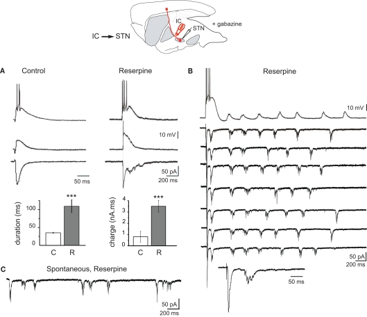
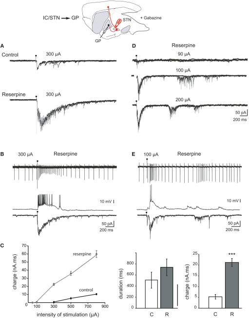
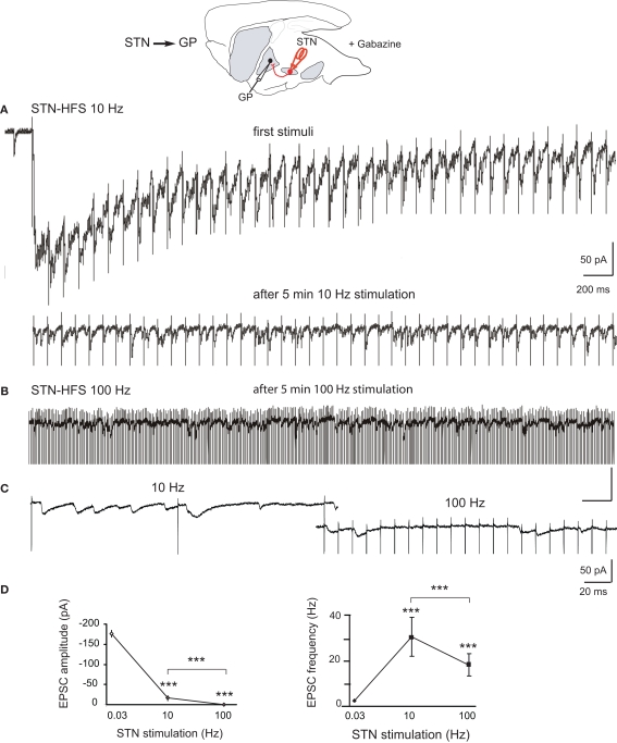
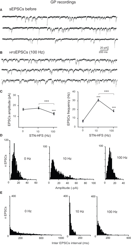
Excessive burst firing in the dopamine-depleted basal ganglia correlates with severe motor symptoms of Parkinson's disease that are attenuated by high frequency electrical stimulation of the subthalamic nucleus (STN). Here we test the hypothesis that pathological bursts in dopamine-deprived basal ganglia are generated within the STN and transmitted to globus pallidus neurons. To answer this question we recorded excitatory synaptic currents and potentials from subthalamic and pallidal neurons in the basal ganglia slice (BGS) from dopamine-depleted mice while continuously blocking GABA(A) receptors. In control mice, a single electrical stimulus delivered to the internal capsule or the rostral pole of the STN evoked a short duration, small amplitude, monosynaptic EPSC in subthalamic neurons. In contrast, in the dopamine-depleted BGS, this monosynaptic EPSC was amplified and followed by a burst of polysynaptic EPSCs that eventually reverberated three to seven times, providing a long lasting response that gave rise to bursts of EPSCs and spikes in GP neurons. Repetitive (10-120 Hz) stimulation delivered to the STN in the dopamine-depleted BGS attenuated STN-evoked bursts of EPSCs in pallidal neurons after several minutes of stimulation but only high frequency (90-120 Hz) stimulation replaced them with small amplitude EPSCs at 20 Hz. We propose that the polysynaptic pathway within the STN amplifies subthalamic responses to incoming excitation in the dopamine-depleted basal ganglia, thereby transforming the STN into a burst generator and entraining pallidal neurons in pathogenic bursting activities. High frequency stimulation of the STN prevents the transmission of this pathological activity to globus pallidus and imposes a new glutamatergic synaptic noise on pallidal neurons.

Keywords: Parkinson; basal ganglia; basal ganglia slice; burst firing; high frequency stimulation; subthalamic nucleus.


Figures

Figure 1
Figure 1
Response of STN neurons to internal capsule stimulation in control and dopamine-depleted BGS in the continuous presence of gabazine (10 μM). Top: schematic illustration of the experimental design with the stimulation (red) and recording sites. (A) Examples of evoked glutamatergic EPSPs (current clamp traces at Vm = −60 mV (top) and Vm = −70 mV (middle) and EPSCs (voltage clamp traces at VH = −70 mV, bottom) recorded from the same STN neuron in response to a single shock (100 μs, 300 μA) delivered to the internal capsule (IC) in the BGS from control (left) and reserpine-treated (right) mice. Note the difference in time calibration between left and right traces. Histograms of the duration and total charge of the IC-evoked STN EPSC recorded in BGS from control (C, white) and reserpine-treated (R, gray) mice (n = 7 and 11, respectively). (B) Example trace of reverberant glutamatergic EPSPs (top trace, Vm = −70 mV) and EPSCs (seven traces, VH = −70 mV) recorded from the same STN neuron in response to a single shock (100 μs, 85 μA) delivered to the internal capsule at 0.03 Hz (arrowhead). The 8th trace is the first part of the voltage clamp response at an expanded time scale to show the early EPSC. (C) Spontaneous bursts of glutamatergic EPSCs (VH = −70 mV) recorded from a STN neuron in a BGS from a reserpine-treated mouse.
Figure 2
Figure 2
Response of GP neurons to stimulation of the internal capsule or rostral STN in control and dopamine-depleted BGS. Top: Schematic illustration of the experimental design with the stimulation (red) and recording sites. (A) Example traces from GP cells recorded from control BGS (top) and BGS from a reserpinized mouse (bottom). A single shock (100 μs, 300 μA) was delivered with the same stimulating electrode to the rostral pole of the STN at the time of the artifact. It evoked compound glutamatergic EPSCs (VH = −70 mV, three superimposed traces). (B) Recordings from the same GP neuron in cell-attached current clamp mode (top), whole-cell current clamp mode (middle, Vm = −60 mV) and whole-cell voltage clamp mode (bottom, VH = −70 mV) in a BGS from a reserpinized mouse. A single shock delivered to the rostral pole of the STN (100 μs, 300 μA) evoked a long lasting train of spikes, an EPSP giving rise to a long lasting train of spikes and a compound EPSC. (C) Total charge (nA.ms) of the compound EPSCs recorded in (A) as a function of the intensity of stimulation (μA; black circles: control, gray circles: reserpine). Histograms of the duration (left), and charge (right) of the compound EPSCs evoked in control (white, C) and reserpine (gray, R) conditions with the same stimulating electrode and at the same intensity of stimulation (300 μA). (D) Reverberating bursts of EPSCs recorded from the same GP neuron in a BGS from a reserpinized mouse. A single shock (100 μs, arrow head) was delivered to the rostral pole of the STN at the indicated intensities. (E) Example trace of a reverberant glutamatergic train of spikes (cell-attached, top), EPSPs (whole-cell, Vm = −60 mV, middle) and bursts of EPSCs (whole-cell, VH = −70 mV, bottom) recorded from the same GP neuron in a BGS from a reserpinized mouse, in response to a single shock (100 μs, 85 μA) delivered to the rostral pole of the STN.
Figure 3
Figure 3
Effects of STN-HFS on the EPSCs evoked in GP neurons in dopamine-depleted BGS. Top: Schematic illustration of the experimental design with the stimulation (red) and recording sites. (A) STN-HFS at 10 Hz evoked EPSCs at 10 Hz. Note that HFS causes an initial shift in holding current that is caused by time-dependent summation of the late components of compound EPSCs. After 5 min of 10 Hz stimulation, the EPSCs evoked in the same GP neuron had a lower amplitude. (B) STN-evoked glutamatergic EPSCs recorded in the same GP neuron as in (A) during STN-HFS at 100 Hz. (C): Close up of traces under 10 and 100 Hz STN-HFS. (D) Averaged data showing the effects of 10 and 100 Hz STN stimulation compared to 0.03 Hz (control) on the amplitude and frequency of the EPSCs evoked in DA-depleted GP neurons. Each data point is the mean ± SEM. For the response at 0.03 Hz we considered the complex response as a whole and plotted its peak amplitude and frequency as if it were a single EPSC.
Figure 4
Figure 4
Comparison of spontaneous glutamatergic EPSCs recorded before and during STN-HFS. (A) Three example traces showing the spontaneous EPSCs (sEPSCs) recorded in voltage clamp mode (VH = −70 mV) from a GP neuron in a BGS from a reserpinized mouse. (B) Three example traces from the same GP neuron as in (A) showing the EPSCs recorded during STN-HFS at 100 Hz (HFSEPSCs, filtered traces without stimulation artifacts). (C) Average data showing the effects of 10 and 100 Hz STN stimulation compared to before HFS (0 Hz) on the amplitude and frequency of the EPSCs recorded in DA-depleted GP neurons. Each data point is the mean ± SEM. Histograms of the amplitudes [bin 1 pA, (D)] and inter EPSCs intervals [bin 10 ms, (E)] for the spontaneous EPSCs (0 Hz) and the HFSEPSCs recorded during 10 or 100 Hz STN stimulation in the same GP neuron, as indicated.

Similar articles

Cited by

References

    1. Ammari R., Lopez C., Bioulac B., Garcia L., Hammond C. (2010). Subthalamic nucleus evokes similar long lasting glutamatergic excitations in pallidal, entopeduncular and nigral neurons in the basal ganglia slice. Neuroscience 166, 808–81810.1016/j.neuroscience.2010.01.011 - DOI - PubMed
    1. Baufreton J., Atherton J. F., Surmeier D. J., Bevan M. D. (2005). Enhancement of excitatory synaptic integration by GABAergic inhibition in the subthalamic nucleus. J. Neurosci. 25, 8505–851710.1523/JNEUROSCI.1163-05.2005 - DOI - PMC - PubMed
    1. Baufreton J., Bevan M. D. (2008). D-2-like dopamine receptor-mediated modulation of activity-dependent plasticity at GABAergic synapses in the subthalamic nucleus. J. Physiol. (Lond.) 586, 2121–2142 - PMC - PubMed
    1. Benabid A. L., Chabardes S., Torres N., Piallat B., Krack P., Fraix V., Pollak P. (2009). Functional neurosurgery for movement disorders: a historical perspective. Prog. Brain Res. 175, 379–39110.1016/S0079-6123(09)17525-8 - DOI - PubMed
    1. Benazzouz A., Gao D. M., Ni Z. G., Piallat B., Bouali-Benazzouz R., Benabid A. L. (2000). Effect of high-frequency stimulation of the subthalamic nucleus on the neuronal activities of the substantia nigra pars reticulata and ventrolateral nucleus of the thalamus in the rat. Neuroscience 99, 289–29510.1016/S0306-4522(00)00199-8 - DOI - PubMed

LinkOut - more resources