

The Role of the spv Genes in Salmonella Pathogenesis
- PMID: 21716657
- PMCID: PMC3117207
- DOI: 10.3389/fmicb.2011.00129
The Role of the spv Genes in Salmonella Pathogenesis
Abstract
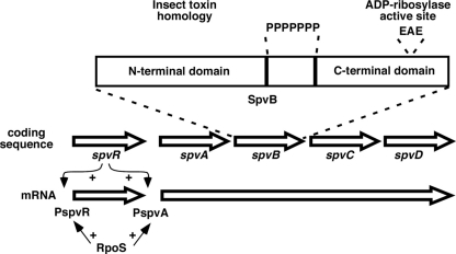
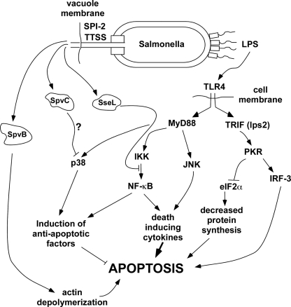
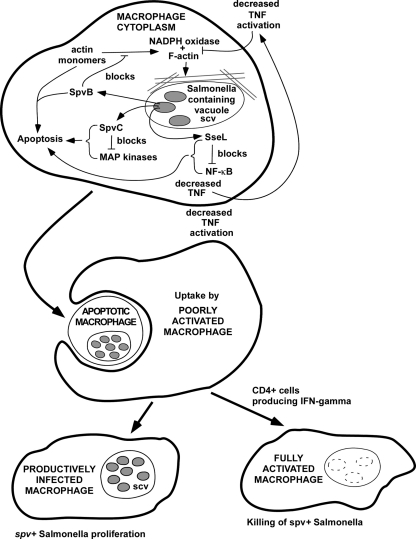
Salmonella strains cause three main types of diseases in people: gastroenteritis, enteric (typhoid) fever, and non-typhoid extra-intestinal disease with bacteremia. Genetic analysis indicates that each clinical syndrome requires distinct sets of virulence genes, and Salmonella isolates differ in their constellation of virulence traits. The spv locus is strongly associated with strains that cause non-typhoid bacteremia, but are not present in typhoid strains. The spv region contains three genes required for the virulence phenotype in mice: the positive transcriptional regulator spvR and two structural genes spvB and spvC. SpvB and SpvC are translocated into the host cell by the Salmonella pathogenicity island-2 type-three secretion system. SpvB prevents actin polymerization by ADP-ribosylation of actin monomers, while SpvC has phosphothreonine lyase activity and has been shown to inhibit MAP kinase signaling. The exact mechanisms by which SpvB and SpvC act in concert to enhance virulence are still unclear. SpvB exhibits a cytotoxic effect on host cells and is required for delayed cell death by apoptosis following intracellular infection. Strains isolated from systemic infections of immune compromised patients, particularly HIV patients, usually carry the spv locus, strongly suggesting that CD4 T cells are required to control disease due to Salmonella that are spv positive. This association is not seen with typhoid fever, indicating that the pathogenesis and immunology of typhoid have fundamental differences from the syndrome of non-typhoid bacteremia.
Keywords: ADP-ribosylation; CD4; Salmonella; apoptosis; non-typhoid bacteremia; phosphothreonine lyase; spv; virulence.
Figures
References
-
- Abrahams G. L., Hensel M. (2006). Manipulating cellular transport and immune responses: dynamic interactions between intracellular Salmonella enterica and its host cells. Cell. Microbiol. 8, 728–737 - PubMed
-
- Allen L. A., DeLeo F. R., Gallois A., Toyoshima S., Suzuki K., Nauseef W. M. (1999). Transient association of the nicotinamide adenine dinucleotide phosphate oxidase subunits p47phox and p67phox with phagosomes in neutrophils from patients with X-linked chronic granulomatous disease. Blood 93, 3521–3530 - PubMed
-
- Arthur G., Nduba V. N., Kariuki S. M., Kimari J., Bhatt S. M., Gilks C. F. (2001). Trends in bloodstream infections among human immunodeficiency virus-infected adults admitted to a hospital in Nairobi, Kenya, during the last decade. Clin. Infect. Dis. 33, 248–256 - PubMed
-
- Blaser M. J., Feldman R. A. (1981). From the centers for disease control. Salmonella bacteremia: reports to the Centers for Disease Control, 1968–1979. J. Infect. Dis. 143, 743–746 - PubMed
Grants and funding
LinkOut - more resources
Full Text Sources
Research Materials