

Severe alterations in lipid composition of frontal cortex lipid rafts from Parkinson's disease and incidental Parkinson's disease
- PMID: 21717034
- PMCID: PMC3188884
- DOI: 10.2119/molmed.2011.00119
Severe alterations in lipid composition of frontal cortex lipid rafts from Parkinson's disease and incidental Parkinson's disease
Abstract
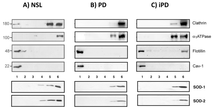
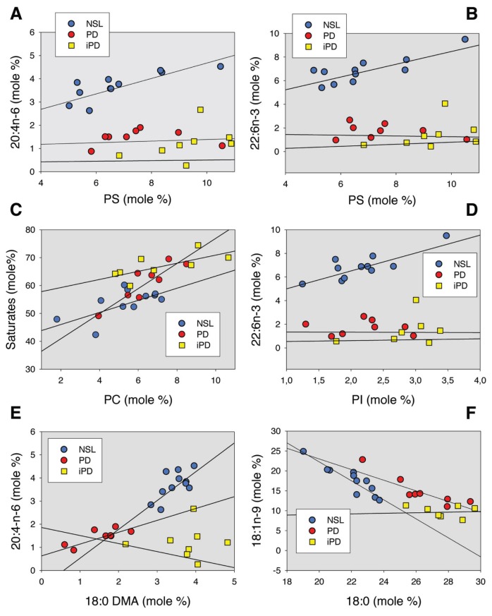
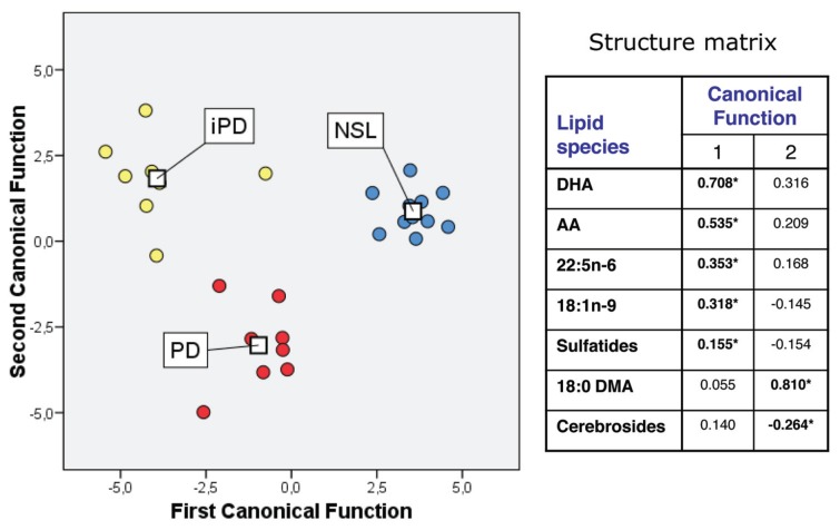
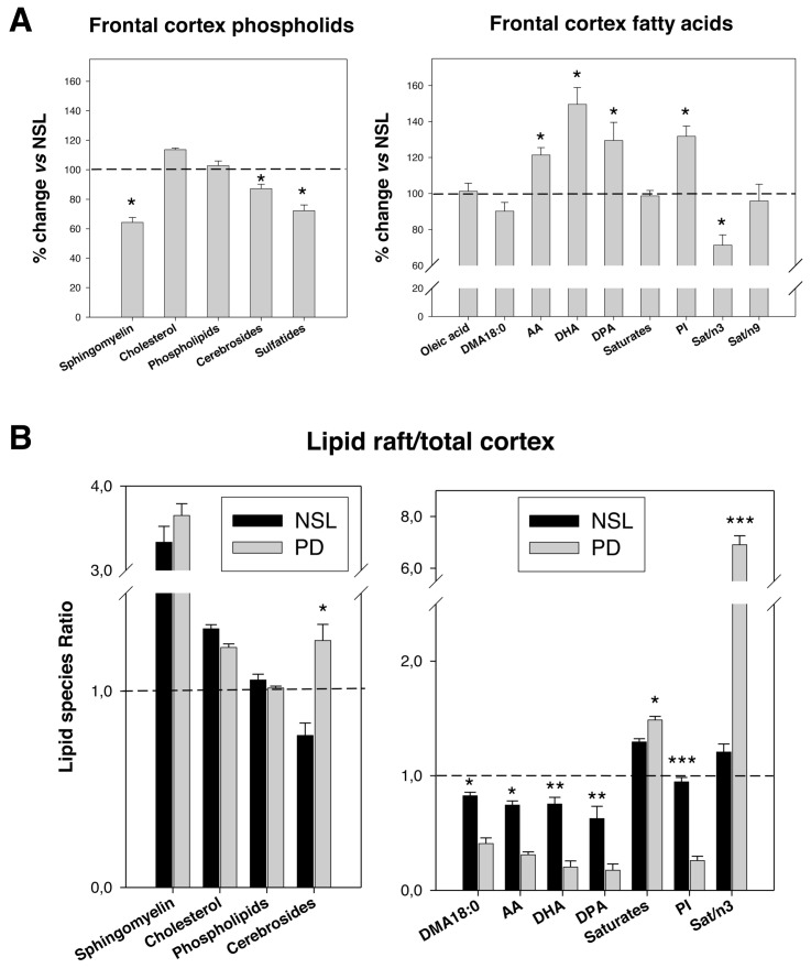
Lipid rafts are cholesterol- and sphingomyelin-enriched microdomains that provide a highly saturated and viscous physicochemical microenvironment to promote protein-lipid and protein-protein interactions. We purified lipid rafts from human frontal cortex from normal, early motor stages of Parkinson's disease (PD) and incidental Parkinson's disease (iPD) subjects and analyzed their lipid composition. We observed that lipid rafts from PD and iPD cortices exhibit dramatic reductions in their contents of n-3 and n-6 long-chain polyunsaturated fatty acids, especially docosahexaenoic acid (22:6-n3) and arachidonic acid (20:4n-6). Also, saturated fatty acids (16:0 and 18:0) were significantly higher than in control brains. Paralleling these findings, unsaturation and peroxidability indices were considerably reduced in PD and iPD lipid rafts. Lipid classes were also affected in PD and iPD lipid rafts. Thus, phosphatidylserine and phosphatidylinositol were increased in PD and iPD, whereas cerebrosides and sulfatides and plasmalogen levels were considerably diminished. Our data pinpoint a dramatic increase in lipid raft order due to the aberrant biochemical structure in PD and iPD and indicate that these abnormalities of lipid rafts in the frontal cortex occur at early stages of PD pathology. The findings correlate with abnormal lipid raft signaling and cognitive decline observed during the development of these neurodegenerative disorders.
Figures
References
-
- Forno LS. Neuropathology of Parkinson’s disease. J Neuropathol Exp Neurol. 1996;55:259–72. - PubMed
-
- Jellinger K, Mizuno Y. Parkinson’s disease. In: Dickson D, editor. Neurodegeneration: The Molecular Pathology of Dementia and Movement Disorders. Basel, Switzerland: ISN Neuropath Press; 2003. pp. 159–87.
-
- Iwatsubo T. Aggregation of α-synuclein in the pathogenesis of Parkinson’s disease. J. Neurol. 2003;250(Suppl 3):11–4. - PubMed
-
- Braak H, del Tredici K. Cortico-basal ganglia-cortical circuitry in Parkinson’s disease reconsidered. Exp Neurol. 2008;212:226–9. - PubMed
-
- Jellinger KA. Lewy body-related synucleinopathy in the aged human brain. J Neural Transm. 2004;111:1219–35. - PubMed
Publication types
MeSH terms
Substances
LinkOut - more resources
Full Text Sources
Other Literature Sources
Medical
Miscellaneous
