Multicenter Study
. 2011 Dec;28(10):781-92.
doi: 10.1055/s-0031-1281509. Epub 2011 Jun 29.

The Stillbirth Collaborative Research Network (SCRN) placental and umbilical cord examination protocol

Affiliations
Multicenter Study

The Stillbirth Collaborative Research Network (SCRN) placental and umbilical cord examination protocol

Halit Pinar et al. Am J Perinatol. 2011 Dec.

Abstract

The Stillbirth Collaborative Research Network (SCRN) was organized to study the scope and causes of stillbirth (SB) in the United States. The objective of this report is to describe the approach used for the placental examination performed as part of the study. The SCRN consists of a multidisciplinary team of investigators from five clinical sites, the National Institute of Child Health and Human Development, and the Data Coordination and Analysis Center. The study is a population-based cohort and nested case-control study, with prospective enrollment of women with SB and live births (LB) at the time of delivery. Detailed and standardized postmortem examination was performed on SB and placental examination in both groups. A total of 663 women with SB and 1932 women with LB were enrolled into the case-control study. In the SB group, there were 707 fetuses. Of these cases, 654 (98.6%) had placental examination. Of these LB controls, 1804 (93.4%) had placental examination. This is the largest prospective study to include population-based SB and LB, using standardized postmortem and placental examination, medical record review, maternal interview, collection of samples, and a multidisciplinary team of investigators collaborating in the analyses. Thus it has the potential to provide high-level evidence regarding the contribution of placental abnormalities to stillbirth.


Figures

Figure 1
Figure 1
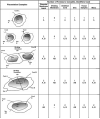
For the purpose of examining various types of placentas consistently, we designed the data extraction forms that could be used in all types of placentation for singleton and multiple gestations. This system considered four elements: (1) umbilical cord(s), (2) placental disc(s), (3) chorioamniotic sac(s) (placental membranes), and (4) dividing membrane(s). The number of umbilical cords was used as a marker for the number of fetuses present in a gestation. In singleton pregnancies, the cord, disc, and sac were all identified as “A.” In multifetal pregnancies, when the delivery personnel marked the cords, the cord ID for the firstborn was designated as “A,” for the second-born as “B,” and so on. If birth order was unknown, cords were arbitrarily designated using the letters M, N, and so on. Identifiers for placental membranes (sacs) were designated with one or more letters (e.g., A, B, AB; M, N, MN) depending on the placentation. Dividing membranes were indicated with two letters (e.g., AB; MN) and placental discs were each indicated with a single letter when discs were distinct and two (or more) letters when fused (with or without a dividing membrane, depending on the number of sacs). Forms for tissue samples and the microscopic examination used the cord ID. For a fused disc, if a dividing membrane could not be observed, an imaginary perpendicular midway between the umbilical cord insertion points was used to distinguish “side A” and “side B” for purposes of sampling and performing the microscopic examination.
Figure 2
Figure 2
Main images taken from the placenta and documentation of sampling. (A) The first image was that of the fetal surface after trimming the membranes and the umbilical cord. If there were any significant membranous or umbilical cord lesions in relation to the placental disc, these were documented before trimming the placental disc. (B) Maternal surface with irregular lobules (cotyledons). (C) Position of the protractor on the maternal surface. (D) Sliced placenta with the umbilical cord marker in place for orientation.
Figure 3
Figure 3
Mock-up of a selection list of random angles and proportional locations for sampling from the placental disc.
Figure 4
Figure 4
This image illustrates the coordinates to be used for the sampling of the placental disc; the same image was repeated after coordinate markers and protractor were added to the placental disc. Frequently, these two images were combined. The maternal surface of the placenta shows the landmarks and placement of the protractor. The placenta is oriented with the long axis horizontal. Locations of the landmarks are marked with pushpins: (1) location of the umbilical cord insertion point (on the fetal side of the placenta); (2A and 2B) leftmost and rightmost borders; (3A and 3B) topmost and bottommost borders. For multiple gestations, additional landmarks were recorded regarding the position of dividing membranes. Coordinates for these landmarks were recorded in the data collection forms. The pins labeled 4A and 4B show the borders marking the randomly assigned sampling axis. The protractor is centered at the landmark corresponding to the location of the umbilical cord insertion point. The white lines indicate the central 2-cm strip, which is used for the histological samples.
Figure 5
Figure 5
Locating random percentages along a sampling ray and its opposite ray. Schematic of the maternal surface of the placenta, viewed from above, showing locations of 0% and 50% on the sampling ray and 40% and 90% on the opposite ray. The location for 0% is slightly beyond the edge of the umbilical cord insertion point (to leave room for a clinical block); 100% is at the margin.
Figure 6
Figure 6
Location of samples relative to the sampling locations and placental slices, overlaid on the diagram in Fig. 5. Schematic of the maternal surface of the placenta, viewed from above. The drawing is to relative scale for a near-term, 22-cm-diameter placenta, assumed to be ~2 cm thick. Specimens for histology (2 × 0.2 cm × full-thickness depth) are cut from the central, 2-cm-wide slice that contains the umbilical cord insertion site. The histological specimens are cut with the 2-cm side perpendicular to the sampling axis. The Stillbirth Collaborative Research Network (SCRN) block is obtained at the sampling location, and the clinical block is collected immediately adjacent to the SCRN block, on the side toward the umbilical cord insertion. An additional SCRN block is cut through the center of the umbilical cord insertion point, and a clinical block is collected immediately adjacent to the SCRN block. Additional slices are made every 1 cm to the edge of the placenta in each direction. SCRN frozen samples are collected preferentially from the adjacent 1-cm slice that is immediately counterclockwise to the first sampling ray. The location point of each frozen sample should line up with the sampling locations whenever possible (there are rules regarding exceptions in certain situations); 1 × 1 cm × full-thickness frozen samples are shown, because this placenta is assumed to be 2-cm thick, yielding ~2 g of placental parenchyma per sample.
Figure 7
Figure 7
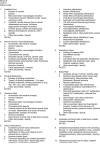
Data elements collected in the placenta microscopic examination. EVT, extravillous trophoblast; LB, liveborn; SB, stillborn; RBC, red blood cells.

References

    1. Driscoll SG, Langston C. College of American Pathologists Conference XIX on the Examination of the Placenta: report of the Working Group on Methods for Placental Examination. Arch Pathol Lab Med. 1991;115:704–708. - PubMed
    1. Kaplan C, Lowell DM, Salafia C. College of American Pathologists Conference XIX on the Examination of the Placenta: report of the Working Group on the Definition of Structural Changes Associated with Abnormal Function in the Maternal/Fetal/Placental Unit in the Second and Third Trimesters. Arch Pathol Lab Med. 1991;115:709–716. - PubMed
    1. The examination of the placenta: patient care and risk management. College of American Pathologists Conference XIX. Northfield, Illinois, September 6–7, 1990. Proceedings. Arch Pathol Lab Med. 1991;115:660–721. - PubMed
    1. Langston C, Kaplan C, Macpherson T, et al. Practice guideline for examination of the placenta: developed by the Placental Pathology Practice Guideline Development Task Force of the College of American Pathologists. Arch Pathol Lab Med. 1997;121:449–476. - PubMed
    1. Committee on Genetics ACOG Committee Opinion No. 383: Evaluation of stillbirths and neonatal deaths. Obstet Gynecol. 2007;110:963–966. - PubMed

Publication types