

Associations of ionizing radiation and breast cancer-related serum hormone and growth factor levels in cancer-free female A-bomb survivors
- PMID: 21718103
- PMCID: PMC3925891
- DOI: 10.1667/rr2631.1
Associations of ionizing radiation and breast cancer-related serum hormone and growth factor levels in cancer-free female A-bomb survivors
Abstract
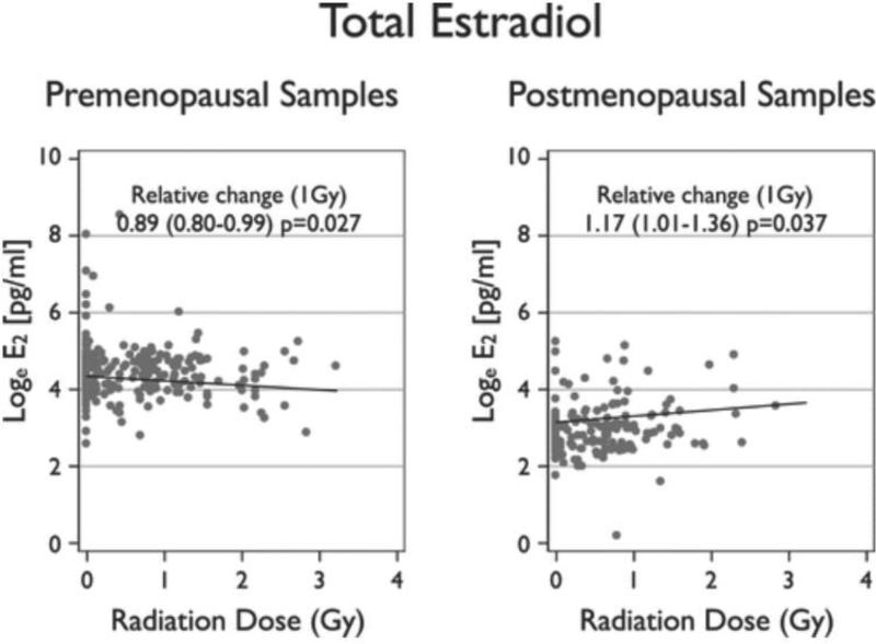
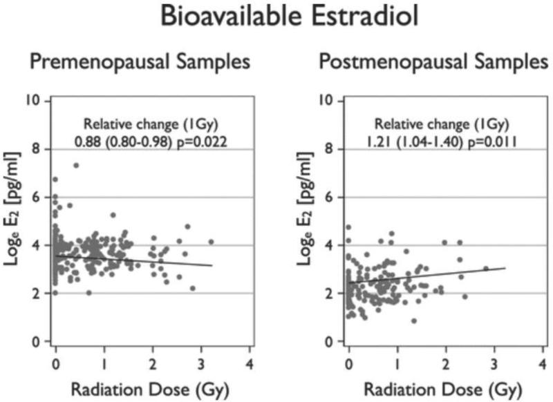
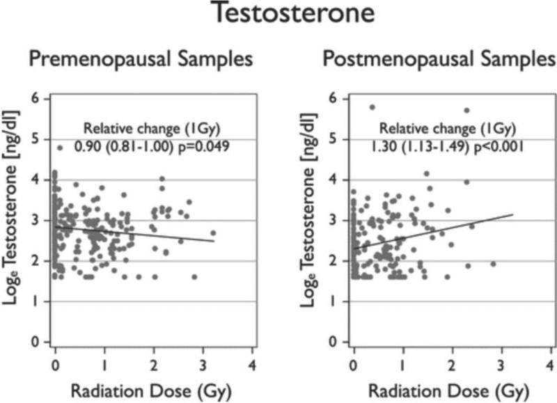
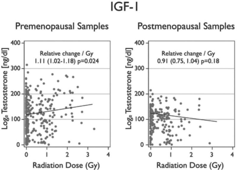
Levels of exposure to ionizing radiation are increasing for women worldwide due to the widespread use of CT and other radiologic diagnostic modalities. Exposure to ionizing radiation as well as increased levels of estradiol and other sex hormones are acknowledged breast cancer risk factors, but the effects of whole-body radiation on serum hormone levels in cancer-free women are unknown. This study examined whether ionizing radiation exposure is associated with levels of serum hormones and other markers that may mediate radiation-associated breast cancer risk. Serum samples were measured from cancer-free women who attended biennial health examinations with a wide range of past radiation exposure levels (N = 412, ages 26-79). The women were selected as controls for separate case-control studies from a cohort of A-bomb survivors. Outcome measures included serum levels of total estradiol, bioavailable estradiol, testosterone, progesterone, prolactin, insulin-like growth factor-1 (IGF1), insulin-like growth factor-binding protein 3 (IGFBP-3), and ferritin. Relationships were assessed using repeated-measures regression models fitted with generalized estimating equations. Geometric mean serum levels of total estradiol and bioavailable estradiol increased with 1 Gy of radiation dose among samples collected from postmenopausal women (17%(1Gy), 95% CI: 1%-36% and 21%(1Gy), 95% CI: 4%-40%, respectively), while they decreased in samples collected from premenopausal women (-11%(1Gy), 95% CI: -20%-1% and -12%(1Gy), 95% CI: -20%- -2%, respectively). Interactions by menopausal status were significant (P = 0.003 and P < 0.001, respectively). Testosterone levels increased with radiation dose in postmenopausal samples (30.0%(1Gy), 95% CI: 13%-49%) while they marginally decreased in premenopausal samples (-10%(1Gy), 95% CI: -19%-0%) and the interaction by menopausal status was significant (P < 0.001). Serum levels of IGF1 increased linearly with radiation dose (11%(1Gy), 95% CI: 2%-18%) and there was a significant interaction by menopausal status (P = 0.014). Radiation-associated changes in serum levels of estradiol, bioavailable estradiol, testosterone and IGF1 were modified by menopausal status at the time of collection. No associations with radiation were observed in serum levels of progesterone, prolactin, IGFBP-3 or ferritin.
Figures
Similar articles
-
Bioavailable serum estradiol may alter radiation risk of postmenopausal breast cancer: a nested case-control study.Int J Radiat Biol. 2018 Feb;94(2):97-105. doi: 10.1080/09553002.2018.1419303. Epub 2018 Jan 16. Int J Radiat Biol. 2018. PMID: 29307255 Free PMC article.
-
Genetic and environmental predictors, endogenous hormones and growth factors, and risk of estrogen receptor-positive breast cancer in Japanese women.Cancer Sci. 2011 Nov;102(11):2065-72. doi: 10.1111/j.1349-7006.2011.02047.x. Epub 2011 Aug 24. Cancer Sci. 2011. PMID: 21790896
-
Premenopausal endogenous steroid hormones and breast cancer risk: results from the Nurses' Health Study II.Breast Cancer Res. 2013 Mar 6;15(2):R19. doi: 10.1186/bcr3394. Breast Cancer Res. 2013. PMID: 23497468 Free PMC article.
-
Cancer risk factors associated with insulin-like growth factor (IGF)-I and IGF-binding protein-3 levels in healthy women: effect modification by menopausal status.Cancer Causes Control. 2009 Dec;20(10):1985-96. doi: 10.1007/s10552-009-9393-0. Cancer Causes Control. 2009. PMID: 19597750
-
The effects of ionizing radiation on domestic dogs: a review of the atomic bomb testing era.Biol Rev Camb Philos Soc. 2021 Oct;96(5):1799-1815. doi: 10.1111/brv.12723. Epub 2021 May 13. Biol Rev Camb Philos Soc. 2021. PMID: 33987930 Free PMC article. Review.
Cited by
-
Sex hormones and risk of breast cancer in premenopausal women: a collaborative reanalysis of individual participant data from seven prospective studies.Lancet Oncol. 2013 Sep;14(10):1009-19. doi: 10.1016/S1470-2045(13)70301-2. Epub 2013 Jul 24. Lancet Oncol. 2013. PMID: 23890780 Free PMC article.
-
Differential effect of parity on rat mammary carcinogenesis after pre- or post-pubertal exposure to radiation.Sci Rep. 2018 Sep 25;8(1):14325. doi: 10.1038/s41598-018-32406-1. Sci Rep. 2018. PMID: 30254198 Free PMC article.
-
Bioavailable serum estradiol may alter radiation risk of postmenopausal breast cancer: a nested case-control study.Int J Radiat Biol. 2018 Feb;94(2):97-105. doi: 10.1080/09553002.2018.1419303. Epub 2018 Jan 16. Int J Radiat Biol. 2018. PMID: 29307255 Free PMC article.
-
Simulated Galactic Cosmic Radiation Exposure-Induced Mammary Tumorigenesis in ApcMin/+ Mice Coincides with Activation of ERα-ERRα-SPP1 Signaling Axis.Cancers (Basel). 2024 Nov 26;16(23):3954. doi: 10.3390/cancers16233954. Cancers (Basel). 2024. PMID: 39682141 Free PMC article.
-
Exposure to ionizing radiation causes long-term increase in serum estradiol and activation of PI3K-Akt signaling pathway in mouse mammary gland.Int J Radiat Oncol Biol Phys. 2012 Oct 1;84(2):500-7. doi: 10.1016/j.ijrobp.2011.12.033. Epub 2012 Feb 28. Int J Radiat Oncol Biol Phys. 2012. PMID: 22381906 Free PMC article.
References
-
- Brenner DJ, Hall EJ. Computed tomography – an increasing source of radiation exposure. N Engl J Med. 2007;357:2277–84. CrossRef, PubMed. - PubMed
-
- Preston DL, Mattsson A, Holmberg E, Shore R, Hildreth NG, Boice JD. Radiation effects on breast cancer risk: a pooled analysis of eight cohorts. Radiat Res. 2002;158:220–35. BioOne, PubMed. - PubMed
-
- Preston DL, Ron E, Tokuoka S, Funamoto S, Nishi N, Soda M, et al. Solid cancer incidence in atomic bomb survivors: 1958–1998. Radiat Res 2007. 168:1–64. BioOne, PubMed. - PubMed
-
- UNSCEAR Sources and effects of ionizing radiation. United Nations Scientific Committee on the Effects of Atomic Radiation: UNSCEAR 2000 Report to the General Assembly, with scientific annexes. 2000.
-
- Hankinson SE, Willett WC, Manson JE, Colditz GA, Hunter DJ, Spiegelman D, et al. Plasma sex steroid hormone levels and risk of breast cancer in postmenopausal women. J Natl Cancer Inst. 1998;90:1292–9. CrossRef, PubMed. - PubMed
Publication types
MeSH terms
Substances
Grants and funding
LinkOut - more resources
Full Text Sources
Medical
Miscellaneous
