. 2012 Feb;165(3):683-92.
doi: 10.1111/j.1476-5381.2011.01568.x.

17(R)-resolvin D1 specifically inhibits transient receptor potential ion channel vanilloid 3 leading to peripheral antinociception

Affiliations

17(R)-resolvin D1 specifically inhibits transient receptor potential ion channel vanilloid 3 leading to peripheral antinociception

S Bang et al. Br J Pharmacol. 2012 Feb.

Abstract

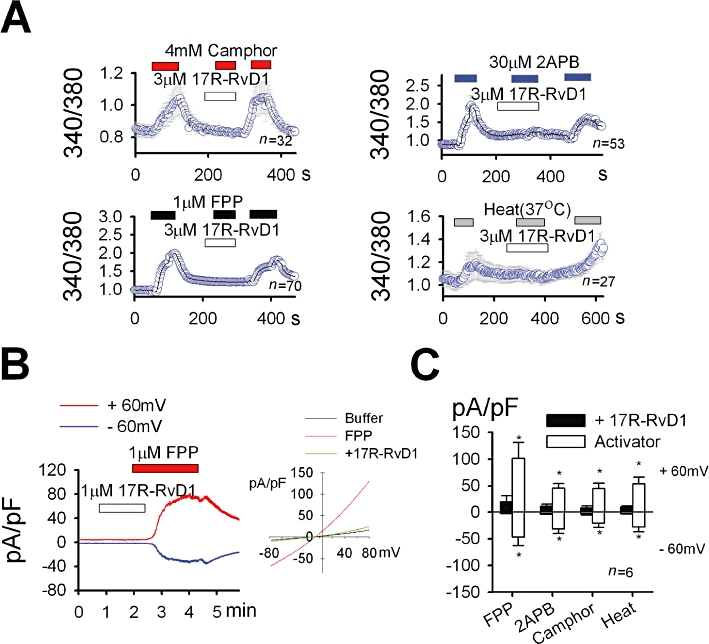
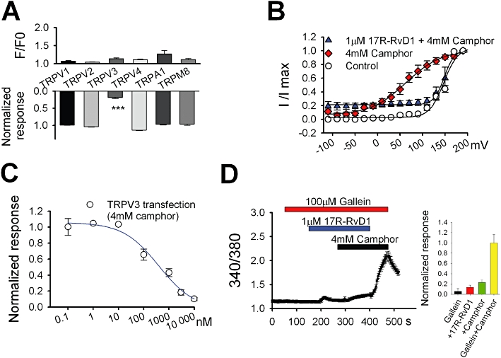
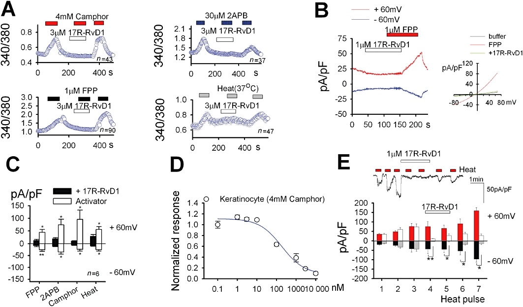
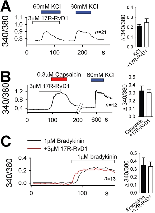
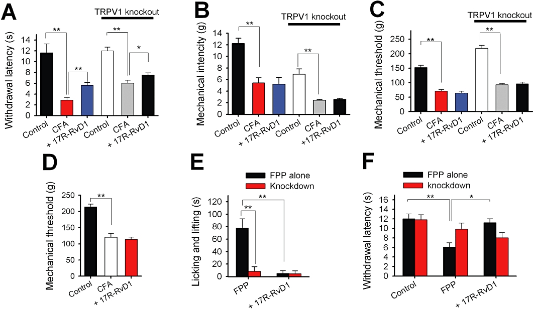
Background and purpose: Transient receptor potential ion channel vanilloid 3 (TRPV3) is expressed in skin keratinocytes and plays an important role in thermal and chemical nociceptions in the periphery. The presence of TRPV3 inhibitors would improve our understanding of TRPV3 function and help to develop receptor-specific analgesics. However, little is known about physiological substances that specifically inhibit TRPV3 activity. Here, we investigated whether 17(R)-resolvin D1 (17R-RvD1), a naturally occurring pro-resolving lipid specifically affects TRPV3 activity.

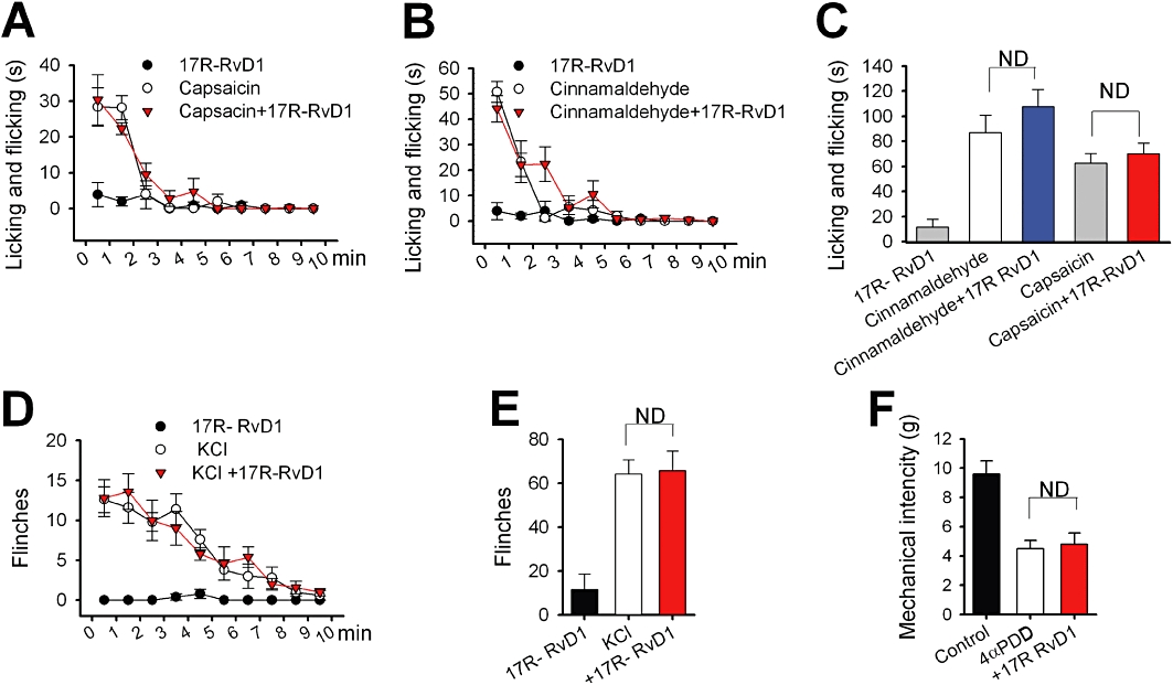
Experimental approach: We examined the effect of 17R-RvD1 on sensory TRP channels using Ca(2+) imaging and whole cell electrophysiology experiments in a HEK cell heterologous expression system, cultured sensory neurons and keratinocytes. We also examined changes in sensory TRP agonist-specific acute licking/flicking or flinching behaviours and mechanical and thermal pain behaviours using Hargreaves, Randall-Selitto and von Frey assay systems in the absence and presence of inflammation.

Key results: We showed that 17R-RvD1 specifically suppresses TRPV3-mediated activity at nanomolar and micromolar concentrations. The voltage-dependence of TRPV3 activation by camphor was shifted rightwards by 17R-RvD1, which indicates its inhibitory mechanism is as a result of a shift in voltage-dependence. Consistently, TRPV3-specific acute pain behaviours were attenuated by locally injected 17R-RvD1. Moreover, the administration of 17R-RvD1 significantly reversed the thermal hypersensitivity that occurs during an inflammatory response. Knockdown of epidermal TRPV3 blunted these antinociceptive effects of 17R-RvD1.

Conclusions and implications: 17R-RvD1 is a novel natural inhibitory substance specific for TRPV3. The results of our behavioural studies suggest that 17R-RvD1 has acute analgesic potential via TRPV3-specific mechanisms.


Figures

Figure 1
Figure 1
17R-RvD1 inhibits TRPV3 activity in the heterologous expression system. (A) 17R-RvD1 attenuated intracellular Ca2+ increases in response to TRPV3 agonists and 37°C heat stimulation. The test concentrations of all three agonists were between their EC50 values and ones that elicit maximal efficacy (Hu et al., 2009; Bang et al., 2010a). (B-C) 17R-RvD1 attenuated current responses to TRPV3 agonists in the whole cell voltage clamp experiments using TRPV3-expressing HEK cells. (B) FPP evoked an outwardly rectifying current increase (n = 7). The current was inhibited by co-application of 1 µM 17R-RvD1. Inset: current–voltage curves for the responses to FPP alone and FPP plus 17R-RvD1 are superimposed. (C) Average current densities induced by TRPV3 activation at ±60 mV are displayed.
Figure 2
Figure 2
Specificity and potency of 17R-RvD1. (A) Changes in intracellular Ca2+ level in cells expressing each TRP were measured during application with 3 µM 17R-RvD1 alone (upper; n = 31–81 for each TRP). The 17R-RvD1 (3 µM)-induced reduction in intracellular Ca2+ increases in response to the agonists was also examined (lower). For each test, the cells were incubated with 17R-RvD1 for 1–3 min. Data for the reduction test were normalized to the averaged responses from each TRP with agonist alone (n = 31–81 for each TRP). (B) Voltage-dependence of TRPV3 activation. The peak inward tail currents at −120 mV from the step protocol were plotted by a function of test voltage steps. Control indicates the data obtained with no drug application. (C) The dose–response curve for 17R-RvD1 on TRPV3 inhibition obtained by Fura-2 Ca2+ imaging. The curve was fitted by the Hill equation. Symbols represent mean values of responses of Ca2+ influx via TRPV3 activation by 4 mM camphor at each 17R-RvD1 dose (n = 17–41). (D) Gallein (100 µM) had no effect on TRPV3 inhibition by 17R-RvD1 in the Fura-2 Ca2+ imaging. Inset: summary of intracellular Ca2+ levels under each drug application normalized to that under 4 mM camphor alone.
Figure 3
Figure 3
17R-RvD1 inhibits TRPV3 activity in keratinocytes. (A) In HaCaT keratinocytes, 17R-RvD1 attenuated intracellular Ca2+ increases in response to TRPV3 agonists and 37°C heat stimulation. (B-C) 17R-RvD1 attenuated current responses to TRPV3 agonists in the whole cell voltage clamp experiments. (B) FPP evoked an outwardly rectifying current increase (n = 7). The current was inhibited by the co-application of 1 µM 17R-RvD1. Inset: current–voltage curves for the responses to FPP alone and FPP plus 17R-RvD1 are superimposed. (C) Average current densities in keratinocyte induced by TRPV3 activation at ±60 mV. (D) The dose–response curve for the inhibitory effect of 17R-RvD1 on 4 mM camphor-induced Ca2+ influx obtained by Fura-2 Ca2+ imaging (n = 17–41). (E) 17R-RvD1 inhibits the TRPV3-mediated response to repeated heat stimulation in the whole cell voltage clamp experiments. Upper: representative current trace for 17R-RvD inhibition at −60 mV. Lower: average current densities in keratinocytes induced by TRPV3 activation at ±60 mV (n = 4–5). During the application of 17R-RvD1 (open and grey columns), the heat responses were decreased by 92.2 ± 9.2% and by 96.8 ± 4.2% at fourth and fifth heat pulses, respectively, compared to the heat responses without 17R-RvD1 (red and black columns) at −60 mV. The final heat responses were increased by 27.8. ± 16.5% and 461.3 ± 138.5% compared to the initial heat responses at −60 mV, with and without 17R-RvD1 addition, respectively.
Figure 4
Figure 4
17R-RvD1 does not affect DRG neuronal responses. In cultured mouse DRG neurons, 17R-RvD1 failed to affect intracellular Ca2+ increases in response to 60 mM KCl-evoked depolarization (A), capsaicin (B) or bradykinin (C). For the bradykinin experiments, different neurons were tested for the 17R-RvD1 effects due to strong desensitization of bradykinin responses. Insets: summary of each stimulation-evoked neuronal Ca2+ influxes with or without 3 µM 17R-RvD1 (n = 21 for KCl, n = 12 for capsaicin and n = 13 for bradykinin).
Figure 5
Figure 5
17R-RvD1 attenuates TRPV3-mediated thermal and chemical nociception. (A) Summary of the changes in hind paw withdrawal latencies upon 17R-RvD1 treatment obtained in Hargreaves assays. The average decrease ratios of the Hargreaves latencies induced by CFA inflammation were 75.3 ± 4.2% (control for wild-type mice, n = 5) and 49.8 ± 5.6% (control for TRPV1-null mice, n = 5). Administration of 17R-RvD1 (30 µM in 20 µL, i.d.) reversed the paw withdrawal latency decreases by 23.5 ± 4.4% (wild type, n = 5) and by 12.2 ± 5.8% (TRPV1-null mice, n = 5) compared with the control withdrawal latencies during inflammation. (B) Summary of the mechanical intensities eliciting hind paw withdrawal in von Frey assays. The average decrease ratios of the von Frey intensities during CFA inflammation were 55.9 ± 7.2% (wild-type mice, n = 6) and 63.5 ± 4.3% (control for TRPV1-null mice, n = 5). Administration of 17R-RvD1 i.d. into the hind paw failed to reverse these decreases in the von Frey intensities (a −1.6 ± 9.5% change compared with the control intensity during inflammation and n = 5 for wild-type mice; a 7.1 ± 9.3% change and n = 5 for TRPV1-null mice). (C–D) Summary of the changes in the mechanical threshold induced by inflammation in Randall-Selitto assays. (C) The average decrease ratios of the mechanical threshold during CFA-induced inflammation were 53.7 ± 3.9% (wild-type mice, n = 6) and 57.1 ± 1.4% (control for TRPV1-null mice, n = 5). Administration of 17R-RvD1 i.d. into the hind paw failed to reverse the decreases in mechanical threshold (a −9.0 ± 8.5% change compared with the control threshold under inflammation; n = 6; a 3.5 ± 8% change and n = 5 for TRPV1-null mice). (D) The average decreased ratios of the mechanical threshold during CFA-induced inflammation were 43 ± 7.4% (rats, n = 5) and i.d. administration of 17R-RvD1 into the hind paw failed to reverse this decrease in mechanical threshold (a −9.4 ± 17% change compared with the control threshold during inflammation; n = 5). (E) A summary of the time spent in licking/lifting behaviours of mice during carrageenan-induced inflammation for 10 min immediately after FPP injection (1 mM in 10 µL). 17R-RvD1 i.d. suppressed FPP-evoked behaviours by 86.6 ± 5.4%. mTRPV3 knockdown with shRNA suppressed FPP-evoked behaviours by 83.4 ± 5.3%, and no further suppression was detected upon the treatment with 17R-RvD1. (F) 17R-RvD1 reversed FPP-induced heat hypersensitivity. In Hargreaves assays, the i.d. FPP treatment significantly reduced Hargreaves latencies by 45.4 ± 5.9% (n = 6). Immediate pretreatment with 17R-RvD1 reversed this reduction (n = 6). mTRPV3 knockdown with shRNA suppressed FPP-induced heat-hypersensitivity (n = 6). No further change in the Hargreaves latencies of the knockdown animals occurred upon the pretreatment with 17R-RvD1 (n = 5). Statistical comparisons were done by anova followed by Bonferroni's post hoc test.
Figure 6
Figure 6
17R-RvD1 failed to affect other related channel-mediated nociceptions. Summaries of the time courses of licking/flicking behaviours in mice treated i.d. with a TRPV1 agonist capsaicin (300 µM in 10 µL) (A), or with a TRPA1 agonist cinnamaldehyde (10 mM in 10 µL) (B), immediately following the injections (n = 5 respectively). Mice were pretreated with 17R-RvD1 (30 µM in 20 µL) immediately before the agonist administrations (n = 5). (C) Summary of the accumulating licking/flicking time of panels (A) and (B). The mean values of the sum of the licking/flicking time during the recording period (10 min) are displayed. (D) Summary of the time course of the flinching behaviours of mice injected intraplantarly with 10 µL KCl solution (140 mM, which evokes sensory neuronal excitation via voltage-gated channel activation) immediately following the injection (n = 5). 17R-RvD1 was administered immediately before the KCl (n = 5). (E) Summary of the total flinching numbers of (D). The mean values of the accumulated flinching numbers during the recording period (10 min) are displayed. (F) 17R-RvD1 failed to reverse a TRPV4 agonist 4-α-phorbol 12,13-didecanoate (4α-PDD)-induced mechanical hypersensitivity. Intraplantar injection with 4α−PDD elicits a decreased von Frey threshold via a TRPV4-specific mechanism (Grant et al., 2007). In our von Frey assays, the i.d. 4α-PDD treatment (1 mM in 10 µL) also significantly reduced von Frey mechanical intensities by 52.2 ± 6.7% (n = 5). Immediate pretreatment with 17R-RvD1 did not significantly affect this reduction (n = 5). Statistical comparisons were done by anova followed by Bonferroni's post hoc test. ND, not significantly different.

References

    1. Alexander SP, Mathie A, Peters JA. Guide to Receptors and Channels (GRAC), 5th edition. Br J Pharmacol. 2011;153(Suppl. 1):S1–S324. - PMC - PubMed
    1. Andersson DA, Gentry C, Moss S, Bevan S. Transient receptor potential A1 is a sensory receptor for multiple products of oxidative stress. J Neurosci. 2008;28:2485–2494. - PMC - PubMed
    1. Ariel A, Serhan CN. Resolvins and protectins in the termination program of acute inflammation. Trends Immunol. 2007;28:176–183. - PubMed
    1. Bandell M, Story GM, Hwang SW, Viswanath V, Eid SR, Petrus MJ, et al. Noxious cold ion channel TRPA1 is activated by pungent compounds and bradykinin. Neuron. 2004;41:849–857. - PubMed
    1. Bang S, Kim KY, Yoo S, Kim YG, Hwang SW. Transient receptor potential A1 mediates acetaldehyde-evoked pain sensation. Eur J Neurosci. 2007a;26:2516–2523. - PubMed

Publication types

MeSH terms