

Consequences of splice variation on Secretin family G protein-coupled receptor function
- PMID: 21718310
- PMCID: PMC3415641
- DOI: 10.1111/j.1476-5381.2011.01571.x
Consequences of splice variation on Secretin family G protein-coupled receptor function
Abstract
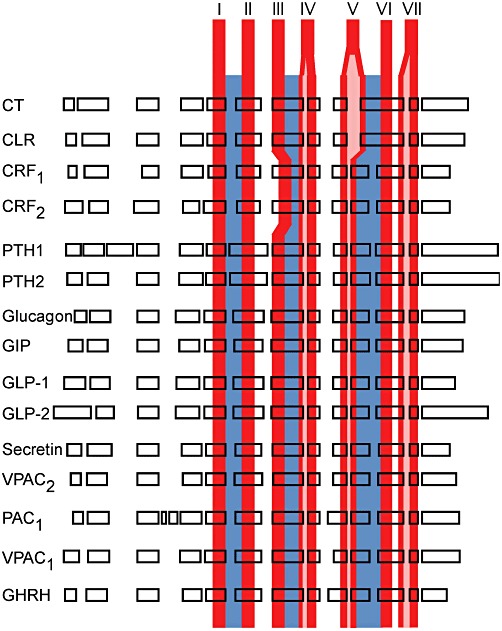
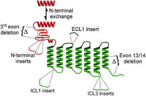
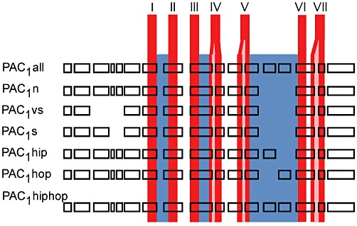
The Secretin family of GPCRs are endocrine peptide hormone receptors that share a common genomic organization and are the subject of a wide variety of alternative splicing. All GPCRs contain a central seven transmembrane domain responsible for transducing signals from the outside of the cell as well as extracellular amino and intracellular carboxyl termini. Members of the Secretin receptor family have a relatively large N-terminus and a variety of lines of evidence support a common mode of ligand binding and a common ligand binding fold. These receptors are best characterized as coupling to intracellular signalling pathways via G(αs) and G(αq) but are also reported to couple to a multitude of other signalling pathways. The intracellular loops are implicated in regulating the interaction between the receptor and heterotrimeric G protein complexes. Alternative splicing of exons encoding both the extracellular N-terminal domain as well as the extracellular loops of some family members has been reported and as expected these splice variants display altered ligand affinity as well as differential activation by endogenous ligands. Various forms of alternative splicing have also been reported to alter intracellular loops 1 and 3 as well as the C-terminus and as one might expect these display differences in signalling bias towards downstream effectors. These diverse pharmacologies require that the physiological role of these splice variants be addressed but should provide unique opportunities for drug design and development.
© 2011 The Authors. British Journal of Pharmacology © 2011 The British Pharmacological Society.
Figures

Similar articles
-
Focus on the splicing of secretin GPCRs transmembrane-domain 7.Trends Biochem Sci. 2009 Sep;34(9):443-52. doi: 10.1016/j.tibs.2009.06.002. Epub 2009 Sep 3. Trends Biochem Sci. 2009. PMID: 19733082 Review.
-
Mapping spatial approximations between the amino terminus of secretin and each of the extracellular loops of its receptor using cysteine trapping.FASEB J. 2012 Dec;26(12):5092-105. doi: 10.1096/fj.12-212399. Epub 2012 Sep 10. FASEB J. 2012. PMID: 22964305 Free PMC article.
-
Fluorescence resonance energy transfer analysis of secretin docking to its receptor: mapping distances between residues distributed throughout the ligand pharmacophore and distinct receptor residues.J Biol Chem. 2007 Nov 9;282(45):32834-43. doi: 10.1074/jbc.M704563200. Epub 2007 Sep 7. J Biol Chem. 2007. PMID: 17827151
-
Signaling mechanisms of secretin receptor.Regul Pept. 2006 Nov 15;137(1-2):95-104. doi: 10.1016/j.regpep.2006.02.011. Epub 2006 Aug 22. Regul Pept. 2006. PMID: 16930743 Review.
-
Ligand binding and activation of the secretin receptor, a prototypic family B G protein-coupled receptor.Br J Pharmacol. 2012 May;166(1):18-26. doi: 10.1111/j.1476-5381.2011.01463.x. Br J Pharmacol. 2012. PMID: 21542831 Free PMC article. Review.
Cited by
-
Neuropeptide G Protein-Coupled Receptors as Oncotargets.Front Endocrinol (Lausanne). 2018 Jun 29;9:345. doi: 10.3389/fendo.2018.00345. eCollection 2018. Front Endocrinol (Lausanne). 2018. PMID: 30008698 Free PMC article. Review.
-
Alternative Splicing of the Pituitary Adenylate Cyclase-Activating Polypeptide Receptor PAC1: Mechanisms of Fine Tuning of Brain Activity.Front Endocrinol (Lausanne). 2013 May 21;4:55. doi: 10.3389/fendo.2013.00055. eCollection 2013. Front Endocrinol (Lausanne). 2013. PMID: 23734144 Free PMC article.
-
Molecular Signature for Receptor Engagement in the Metabolic Peptide Hormone Amylin.ACS Pharmacol Transl Sci. 2018 Apr 23;1(1):32-49. doi: 10.1021/acsptsci.8b00002. eCollection 2018 Sep 14. ACS Pharmacol Transl Sci. 2018. PMID: 32219203 Free PMC article.
-
CGRP physiology, pharmacology, and therapeutic targets: migraine and beyond.Physiol Rev. 2023 Apr 1;103(2):1565-1644. doi: 10.1152/physrev.00059.2021. Epub 2022 Dec 1. Physiol Rev. 2023. PMID: 36454715 Free PMC article. Review.
-
The role of GPCRs in bone diseases and dysfunctions.Bone Res. 2019 Jul 8;7:19. doi: 10.1038/s41413-019-0059-6. eCollection 2019. Bone Res. 2019. PMID: 31646011 Free PMC article. Review.
References
-
- Albrandt K, Mull E, Brady EM, Herich J, Moore CX, Beaumont K. Molecular cloning of two receptors from rat brain with high affinity for salmon calcitonin. FEBS Lett. 1993;325:225–232. - PubMed
-
- Ardati A, Goetschy V, Gottowick J, Henriot S, Valdenaire O, Deuschle U, et al. Human CRF2 alpha and beta splice variants: pharmacological characterization using radioligand binding and a luciferase gene expression assay. Neuropharmacology. 1999;38:441–448. - PubMed
-
- Bazarsuren A, Grauschopf U, Wozny M, Reusch D, Hoffmann E, Schaefer W, et al. In vitro folding, functional characterization, and disulfide pattern of the extracellular domain of human GLP-1 receptor. Biophys Chem. 2002;96:305–318. - PubMed
-
- Bergwitz C, Gardella TJ, Flannery MR, Potts JT, Kronenberg HM, Goldring SR, et al. Full activation of chimeric receptors by hybrids between parathyroid hormone and calcitonin. Evidence for a common pattern of ligand-receptor interaction. J Biol Chem. 1996;271:26469–26472. - PubMed
Publication types
MeSH terms
Substances
LinkOut - more resources
Full Text Sources
