. 2011 Aug;179(2):795-806.
doi: 10.1016/j.ajpath.2011.04.015. Epub 2011 Jun 12.

Pathogenic relevance of IgG and IgM antibodies against desmoglein 3 in blister formation in pemphigus vulgaris

Affiliations

Pathogenic relevance of IgG and IgM antibodies against desmoglein 3 in blister formation in pemphigus vulgaris

Kazuyuki Tsunoda et al. Am J Pathol. 2011 Aug.

Abstract

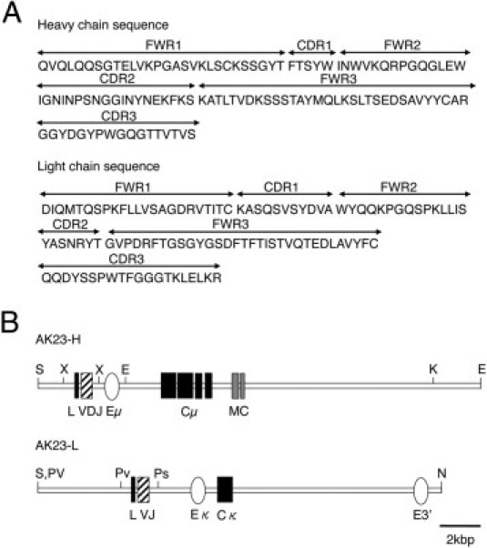
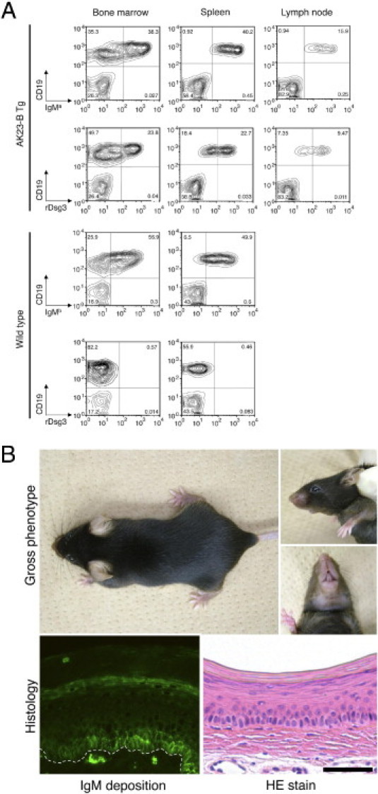
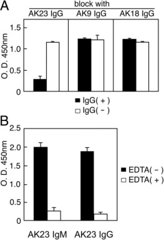
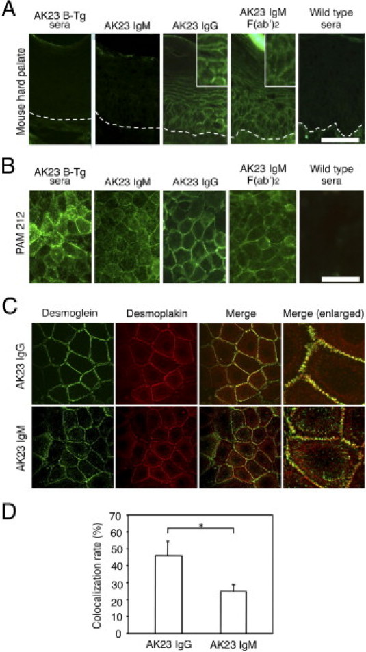
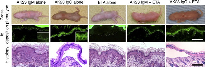
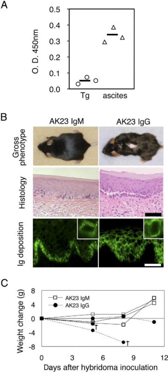
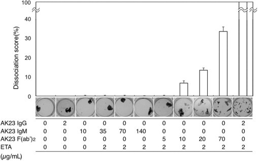
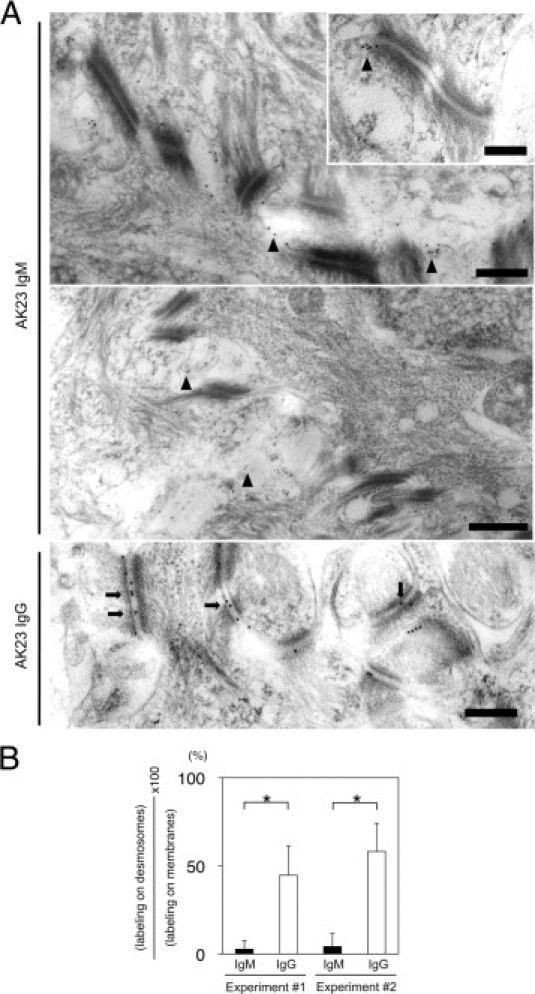
Pemphigus vulgaris is an autoimmune disease caused by IgG antibodies against desmoglein 3 (Dsg3). Previously, we isolated a pathogenic mAb against Dsg3, AK23 IgG, which induces a pemphigus vulgaris-like phenotype characterized by blister formation. In the present study, we generated a transgenic mouse expressing AK23 IgM to examine B-cell tolerance and the pathogenic role of IgM. Autoreactive transgenic B cells were found in the spleen and lymph nodes, whereas anti-Dsg3 AK23 IgM was detected in the cardiovascular circulation. The transgenic mice did not develop an obvious pemphigus vulgaris phenotype, however, even though an excess of AK23 IgM was passively transferred to neonatal mice. Similarly, when hybridoma cells producing AK23 IgM were inoculated into adult mice, no blistering was observed. Immunoelectron microscopy revealed IgM binding at the edges of desmosomes or interdesmosomal cell membranes, but not in the desmosome core, where AK23 IgG binding has been frequently detected. Furthermore, in an in vitro dissociation assay using cultured keratinocytes, AK23 IgG and AK23 IgM F(ab')(2) fragments, but not AK23 IgM, induced fragmentation of epidermal sheets. Together, these observations indicate that antibodies must gain access to Dsg3 integrated within desmosomes to induce the loss of keratinocyte cell-cell adhesion. These findings provide an important framework for improved understanding of B-cell tolerance and the pathophysiology of blister formation in pemphigus.


Figures

Figure 1
Figure 1
Structures of the AK23 H and L immunoglobulin transgene constructs. A: The amino acid sequences of the H and L chains of AK23 were determined from the DNA sequence of AK23 single-chain variable-region fragment. The FWR and CDR domains were annotated accordingly.B: The AK23 heavy-chain variable region (contained in an XbaI-XbaI fragment) was linked with an H-chain promoter and a μ-isotype constant region (Cμ) that included a transmembrane domain (MC). The AK23 L-chain (κ isotype) variable region (contained in a PvuII-PstI fragment was linked to a κ-chain promoter and constant region. E, EcoRI; Eμ, μ-region enhancer; Eκ, κ-region enhancer; K, KpnI; N, NotI; Ps, PstI; Pv, PvuII; S, SalI; X, XbaI.
Figure 2
Figure 2
Dsg3-specific AK23 IgM transgenic B cells escape tolerance. A: Fluorescence-activated cell sorting analysis of AK23 IgM transgenic mice detected IgMa-positive B cells in lymphoid organs, including bone marrow (38.3%), spleen (40.2%), and lymph nodes (15.9%). Dsg3-reactive IgM-expressing B cells were also detected in bone marrow (23.8%), spleen (22.7%), and lymph nodes (9.47%). In wild-type mice, IgMb-positive B cells, but not Dsg3-reactive B cells, were detected in lymphoid organs. B: Macroscopic and microscopic phenotype of AK23 IgM transgenic mice. AK23 IgM transgenic mice exhibited no obvious PV-like characteristics, such as patchy hair loss or weight loss. Although IgM was found deposited on the cell surfaces of basal layer keratinocytes in vivo, no blisters were formed. The dotted line indicates the basement membrane. Scale bar = 50 μm.
Figure 3
Figure 3
AK23 IgM recognizes the same Dsg3 epitope as AK23 IgG. A: The binding of AK23 IgM in a mouse Dsg3 ELISA was significantly blocked by AK23 IgG, but not by AK9 IgG or AK18 IgG. IgG(+), with AK IgG pretreatment; IgG(−), without AK IgG pretreatment. B: AK23 IgM reactivity was tested by mouse Dsg3 ELISA performed with and without EDTA (5 mmol/L) pretreatment.
Figure 4
Figure 4
Comparisons of the immunoreactivities of AK23 IgM, AK23 IgG, and F(ab′)2 fragments of AK23 IgM. A: Indirect immunofluorescence (IIF) staining of hard palate tissue from wild-type mice. Sera from AK23 IgM transgenic mice and AK23 IgM mAb yielded no signal, whereas AK23 IgG and AK23 IgM F(ab′)2 yielded linear cell surface staining. Insets, digitally enlarged. Dotted lines indicate the basement membrane. B: Live staining of mouse PAM212 keratinocytes. Both AK23 IgM and AK23 IgG stained keratinocyte cell surfaces, but the staining patterns were quite different. AK23 IgM appeared as coarse dots on cell border-free areas. C: AK23 IgM and AK23 IgG (green) were costained with desmoplakin (red). Although AK23 IgG largely colocalized with desmoplakin, AK23 IgM did not. D: Quantification of colocalization indicated significantly lower colocalization of DP with AK23 IgM than with AK23 IgG. *P < 0.005. Scale bars = 50 μm.
Figure 5
Figure 5
Evaluation of the pathogenic activity of AK23 IgM by passive transfer assay using neonatal mice. Neonatal mice injected with AK23 IgG (150 μg), but not AK23 IgM (150 μg), developed microscopic blisters. The staining pattern of in vivo bound AK23 IgM was characterized by low-intensity coarse dots, whereas that of AK23 IgG was linear. Mice in neither treatment group developed macroscopic blisters, because of functional compensation by Dsg1 in the skin. When coinjected with half of the minimum effective dose of exfoliative toxin A (ETA), a Dsg1-specific serine protease produced by Staphylococcus aureus, AK23 IgG induced the formation of blisters and yielded typical PV histological features, whereas AK23 IgM produced no obvious signs of blister formation. Dotted lines indicate basement membrane. Scale bar = 50 μm (insets, digitally enlarged).
Figure 6
Figure 6
Evaluation of the pathogenic activity of AK23 IgM by ascites formation assay using adult mice. A: Circulating AK23 IgM titers were approximately six times higher in mice inoculated with AK23 IgM-producing hybridoma cells (triangles) than in AK23 IgM transgenic mice (circles). Sera were diluted 1:5 and analyzed by mouse Dsg3 ELISA. B: Phenotypes of mice injected with AK23 IgM- and AK23 IgG-producing hybridoma cells. Although mice injected with AK23 IgG hybridoma cells exhibited patchy hair loss and suprabasal acantholysis, those that received AK23 IgM hybridoma cells developed no apparent PV phenotype. Although both mice displayed in vivo deposition of AK23 antibodies, IgM was essentially restricted to the basal cell layer of the oral mucosa, appearing as coarse dots, whereas IgG strongly and evenly stained the cell surfaces of keratinocytes in multiple mucosal layers. Images are representative of 18 recipient mice that received AK23 IgM hybridoma cells. Insets, digitally enlarged. C: Changes in body weight were monitored after the injection of hybridoma cells, because the development of oral erosions has the potential to impair food intake. Whereas body weight increased in mice injected with AK23 IgM hybridoma cells, it declined in animals injected with AK23 IgG hybridoma cells, and some of the animals died (n = 3). Scale bars = 50 μm. O.D., optical density; Tg, AK23 IgM transgenic; ascites, mice with AK23 IgM hybridoma cells; †, day of death.
Figure 7
Figure 7
Quantitative evaluation of the pathogenic activity of AK23 IgM by in vitro dissociation assay using cultured human epidermal keratinocytes. To quantitatively evaluate the pathogenic activity of AK23 IgM, we performed an in vitro dissociation assay using NHEK cells. Relative dissociation scores were calculated relative to the dissociation score for AK23 IgG plus ETA. Whereas AK23 IgM exhibited no dissociation activity at concentrations of 35 or 140 μg/mL, AK23 IgM F(ab′)2 fragments yielded dissociation scores of >6.7% and >33.3% at concentrations of 10.0 and 70.0 μg/mL, respectively. Doses of monoclonal antibodies and of ETA are indicated along the x axis.
Figure 8
Figure 8
Ultrastructural localization of AK23 IgM and AK23 IgG in vivo in hard palate of adult mice receiving hybridoma cells. A: AK23 IgM was deposited largely to the cell membranes between desmosomes or the edges of desmosomes in vivo (arrowheads), but not in the desmosome core. Inset, representative image of AK23 IgM deposition. AK23 IgG was heavily deposited in the midlines of desmosomes (arrows). B: Quantitative analysis indicates significantly stronger labeling of desmosomes in mice injected with AK23 IgG-producing hybridoma cells than in those injected with AK23 IgM-producing cells. Scale bars: 500 nm (main images); 200 nm (inset). *P < 0.01, t-test.

References

    1. Amagai M., Klaus-Kovtun V., Stanley J.R. Autoantibodies against a novel epithelial cadherin in pemphigus vulgaris, a disease of cell adhesion. Cell. 1991;67:869–877. - PubMed
    1. Amagai M. Pemphigus. In: Bolognia J.L., Jorizzo J.L., Rapini R.P., editors. ed 2. vol 1. Mosby Elsevier; London: 2008. pp. 417–429. (Dermatology).
    1. Amagai M., Karpati S., Prussick R., Klaus-Kovtun V., Stanley J.R. Autoantibodies against the amino-terminal cadherin-like binding domain of pemphigus vulgaris antigen are pathogenic. J Clin Invest. 1992;90:919–926. - PMC - PubMed
    1. Amagai M., Hashimoto T., Shimizu N., Nishikawa T. Absorption of pathogenic autoantibodies by the extracellular domain of pemphigus vulgaris antigen (Dsg3) produced by baculovirus. J Clin Invest. 1994;94:59–67. - PMC - PubMed
    1. Tsunoda K., Ota T., Aoki M., Yamada T., Nagai T., Nakagawa T., Koyasu S., Nishikawa T., Amagai M. Induction of pemphigus phenotype by a mouse monoclonal antibody against the amino-terminal adhesive interface of desmoglein 3. J Immunol. 2003;170:2170–2178. - PubMed

Publication types

MeSH terms