Case Reports
. 2011 May 12;16(5):213-6.
doi: 10.1186/2047-783x-16-5-213.

Intrahepatic type II gall bladder perforation by a gall stone in a CAPD patient

Affiliations
Case Reports

Intrahepatic type II gall bladder perforation by a gall stone in a CAPD patient

T Göbel et al. Eur J Med Res. .

Abstract

Introduction: Perforation of the gall bladder represents a rare, but life-threatening complication of cholecystitis. Clinical presentation may vary between severe peritonism in acute perforation and absence of symptoms in subacute or chronic progression of perforation. Abdominal imaging like ultrasound or CT-scan are important tools for immediate diagnose of gall bladder perforation.

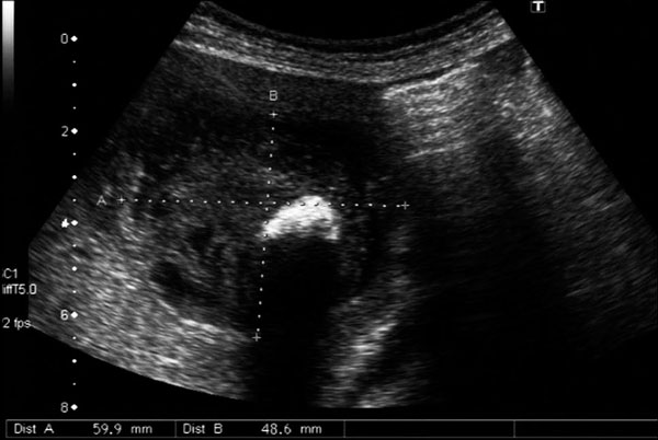
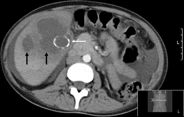
Case presentation: We report a case of a 30-year old female patient with end-stage kidney disease treated by continuous ambulatory peritoneal dialysis (CAPD) who was admitted to the emergency room with fever and mild abdominal pain. A type II gall bladder perforation by a solitary gall stone with development of a liver abscess was detected by abdominal ultrasound.

Conclusion: Gall bladder perforations are rare but have to be considered in patients with abdominal pain and fever. Abdominal ultrasound is a reliable tool to establish diagnosis.


Figures

Figure 1
Figure 1
Large solitary concrement of the gall bladder with a maximal diameter of 27 mm, five years before onset of the gall bladder perforation.
Figure 2
Figure 2
Intrahepatic abscess with dislocation of the solitary concrement from the perforated gall bladder into the abscess cavity.
Figure 3
Figure 3
CT-scan correlating to US-images (see figure 2): The gall stone (white arrow) presents as a circular hyperdense structure within the intrahepatic abscess cavity (black arrows). Free intraabdominal fluid in continuous peritoneal dialysis.
Figure 4
Figure 4
Coronary secondary reconstruction of CT-scan in axial phase. The intrahepatic abscess presents with hyperdense margins (black arrow). Clear illustration which accentuates the extent of the abscess and intrahepatic location of the partial calcified gall stone (white arrow). Free intraabdominal fluid with distal end of the peritoneal catheter in right lower abdomen (white arrowhead).
Figure 5
Figure 5
Follow-up sonography of liver abscess area. A: 8 days after operation (46 × 37 mm), B: 34 days after operation (46 × 25 mm), C: 68 days after operation (21 × 19 mm).

References

    1. Wallace R, Allen A. Acute cholecystitis. Arch Surg. 1943;43(5):762–72.
    1. MacDonald JA. Perforation of the gallbladder associated with acute cholecystitis: 8-year review of 20 cases. Ann surg. 1966;164(5):849–52. doi: 10.1097/00000658-196611000-00010. - DOI - PMC - PubMed
    1. Larmi TK, Kairaluoma MI, Junila J, Laitinen S, Stahlberg M, Fock HG. Perforation of the gallbladder. A retrospective comparative study of cases from 1946-1956 and 1969-1980. Acta Chir Scand. 1984;150(7):557–60. - PubMed
    1. Stefanidis D, Sirinek KR, Bingener J. Gallbladder perforation: risk factors and outcome. J Surg Res. 2006;131(2):204–8. doi: 10.1016/j.jss.2005.11.580. - DOI - PubMed
    1. Kiviluoto T, Siren J, Luukkonen P, Kivilaakso E. Randomised trial of laparoscopic versus open cholecystectomy for acute and gangrenous cholecystitis. Lancet. 1998;351(9099):321–5. doi: 10.1016/S0140-6736(97)08447-X. - DOI - PubMed

Publication types