. 2011 Oct;134(Pt 10):2982-3010.
doi: 10.1093/brain/awr129. Epub 2011 Jun 29.

Dravet syndrome as epileptic encephalopathy: evidence from long-term course and neuropathology

Affiliations

Dravet syndrome as epileptic encephalopathy: evidence from long-term course and neuropathology

Claudia B Catarino et al. Brain. 2011 Oct.

Abstract

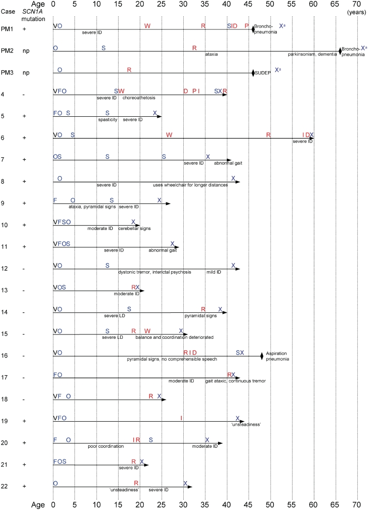
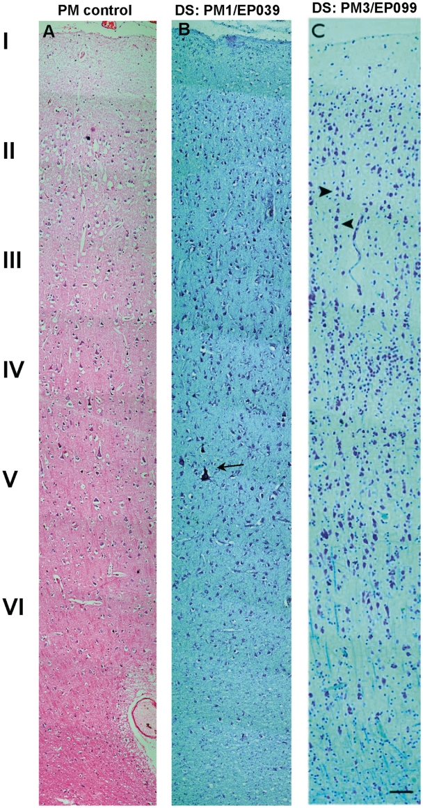
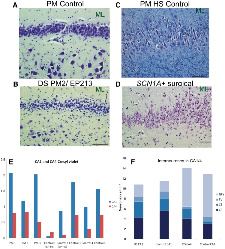
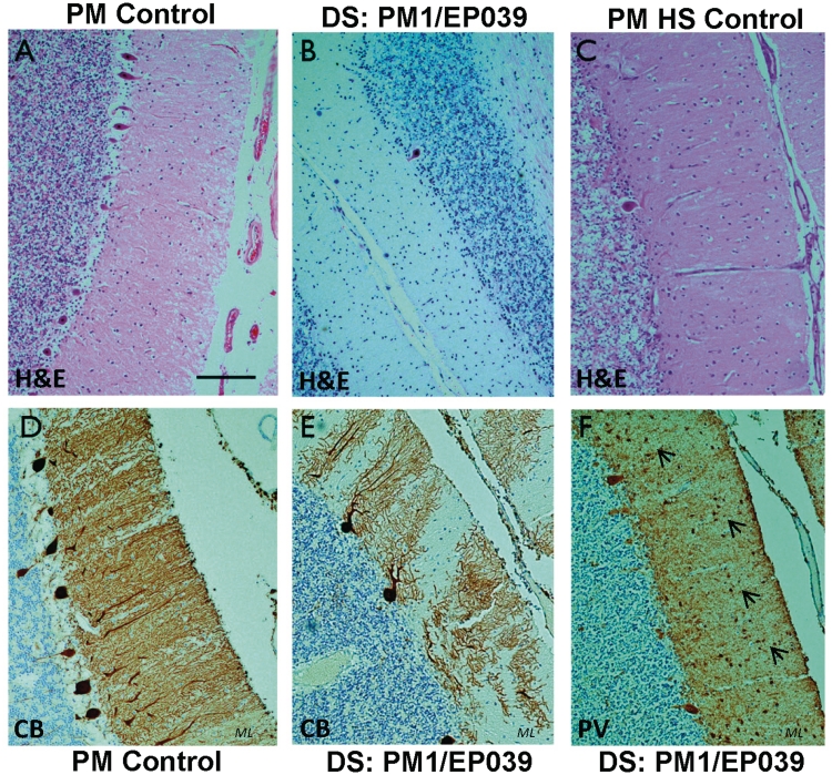
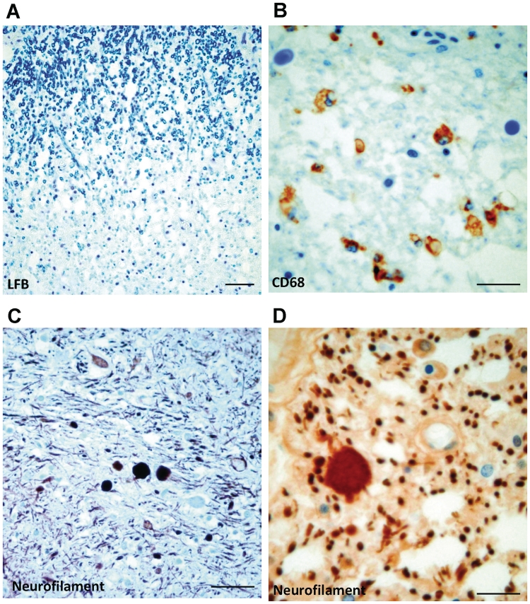
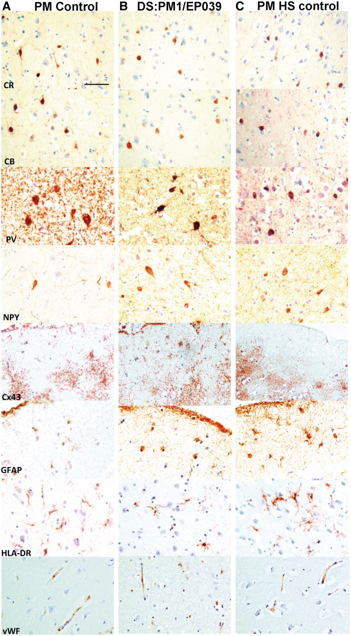
Dravet syndrome is an epilepsy syndrome of infantile onset, frequently caused by SCN1A mutations or deletions. Its prevalence, long-term evolution in adults and neuropathology are not well known. We identified a series of 22 adult patients, including three adult post-mortem cases with Dravet syndrome. For all patients, we reviewed the clinical history, seizure types and frequency, antiepileptic drugs, cognitive, social and functional outcome and results of investigations. A systematic neuropathology study was performed, with post-mortem material from three adult cases with Dravet syndrome, in comparison with controls and a range of relevant paediatric tissue. Twenty-two adults with Dravet syndrome, 10 female, were included, median age 39 years (range 20-66). SCN1A structural variation was found in 60% of the adult Dravet patients tested, including one post-mortem case with DNA extracted from brain tissue. Novel mutations were described for 11 adult patients; one patient had three SCN1A mutations. Features of Dravet syndrome in adulthood include multiple seizure types despite polytherapy, and age-dependent evolution in seizure semiology and electroencephalographic pattern. Fever sensitivity persisted through adulthood in 11 cases. Neurological decline occurred in adulthood with cognitive and motor deterioration. Dysphagia may develop in or after the fourth decade of life, leading to significant morbidity, or death. The correct diagnosis at an older age made an impact at several levels. Treatment changes improved seizure control even after years of drug resistance in all three cases with sufficient follow-up after drug changes were instituted; better control led to significant improvement in cognitive performance and quality of life in adulthood in two cases. There was no histopathological hallmark feature of Dravet syndrome in this series. Strikingly, there was remarkable preservation of neurons and interneurons in the neocortex and hippocampi of Dravet adult post-mortem cases. Our study provides evidence that Dravet syndrome is at least in part an epileptic encephalopathy.


Figures

Figure 1
Figure 1
Timelines in Dravet syndrome—milestones in disease evolution. D = dysphagia; F = febrile seizure; I = incontinence; ID = intellectual disability; np = not possible; O = onset of afebrile seizures; P = percutaneous endoscopic gastrostomy (PEG); R = residential care; S = status epilepticus; SUDEP = sudden unexplained death in epilepsy; V = vaccination; X = diagnosis; W = wheelchair-dependent; a = diagnosis made after death; black diamond = death; horizontal arrow = living patient; + = SCN1A change found; − = no SCN1A change found.
Figure 2
Figure 2
Brain MRI findings in adults with Dravet syndrome and SCN1A mutation. Cerebellar atrophy (A, sagittal T1, Case 6) was a feature in some cases. Case 21 was the only adult case with Dravet syndrome in our series with hippocampal sclerosis (left in this case) evident on MRI (B, coronal T2). Case 6 had a stereotactic thalamotomy at the age of 16 years (C, sagittal T1 and D, coronal T2). Arrows show the location of the main abnormalities in each image.
Figure 3
Figure 3
EEG findings. For Case 6, routine EEG showing background of bilateral diffuse slow activity at 3–5 Hz, and very rare low amplitude sharp waves/spikes, more apparent in frontal regions, right > left (A, bipolar montage). For Case 5, video–EEG telemetry at the age of 26 years, showed bihemispheric cortical dysfunction and bifrontal interictal epileptiform discharges (B, bipolar longitudinal montage). Several complex motor seizures were recorded, some with non-lateralized frontocentral EEG onset (C, combined longitudinal and transverse bipolar montage). Electrographic seizures were also recorded with right posterior temporal pattern (D, bipolar longitudinal montage).
Figure 4
Figure 4
Frontal cortex—histological staining. (A) Haematoxylin and eosin shows the normal frontal cortex from a post-mortem control with no known neurological disease. (B) Cresyl violet shows the motor cortex of the adult Dravet syndrome (DS) case, PM1/EP039, with good preservation of the cortical laminae and Betz cells (arrow). (C) Cresyl violet and Luxol fast blue show the frontal cortex from the adult post-mortem Dravet syndrome case, PM3/EP099, with a focal ‘micro-columnar’ appearance (arrowheads to columnar alignment). Haematoxylin and eosin-stained section is 7 µm thick while Luxol fast blue and cresyl violet-stained sections are 14 µm. Scale bar = 100 µm.
Figure 5
Figure 5
Hippocampus, histological staining and interneuronal cell counts. Cresyl violet shows the normal hippocampus from a post-mortem (PM) control with no known neurological disease (A), and the adult post-mortem case with Dravet syndrome (DS), PM2/EP213 (B). In contrast, pyramidal cell loss in the left cornu ammonis-4 and granule cell dispersion are seen in the hippocampal sclerosis post-mortem (PM HS) control (C), and the SCN1A+ surgical case (D). (E) Stereological quantification of cresyl violet-stained neurons shows lower numbers of pyramidal cells in cornu ammonis-1 and -4 for hippocampal sclerosis post-mortem controls (Control 1 and 2 EP-HS) compared with adult post-mortem cases with Dravet syndrome (PM1–3) and post-mortem controls with no known neurological disease (Controls 3–5). (F) Areal 2D counts of calbindin (CB), calretinin (CR), parvalbumin (PV) and neuropeptide Y (NPY)-immunopositive cells in the cornu ammonis-1 and -4 show that the average number of hippocampal interneurons in the adult post-mortem Dravet syndrome (n = 3) and controls with no known neurological disease (n = 2) is not markedly different. Refer to Fig. 10 (hippocampus immunolabelling) for images of calbindin, calretinin, parvalbumin and neuropeptide Y immunoreactivities in the hippocampus of cases with Dravet syndrome and controls. Scale bar = 50 µm. CA = cornu ammonis; ML = molecular layer.
Figure 6
Figure 6
Cerebellum, histological staining and immunolabelling. (A) Haematoxylin and eosin (H&E) shows a normal cerebellum from a post-mortem control with no known neurological disease. The same stain shows Purkinje cell loss in the cerebellum of the adult post-mortem Dravet syndrome case, PM1/EP039 (B), and a hippocampal sclerosis post-mortem control (C). The loss of Purkinje cells and their processes, which normally extend into the molecular layer as observed in D, is evident in calbindin- and parvalbumin-immunolabelled cerebellar sections from the case with Dravet syndrome, PM1/EP039 (E and F). Small, parvalbumin-immunopositive cells are still observed in the molecular layer of the Dravet syndrome cerebellum (F, arrows). Scale bar = 100 µm. ML = molecular layer.
Figure 7
Figure 7
Brainstem and spinal cord—histological staining and immunolabelling. (A) Luxol fast blue (LFB) section shows a cord area with myelin pallor in the dorsal column of the adult post-mortem Dravet syndrome case, PM1/EP039, where no myelin debris is observed. (B) The same area immunolabelled with the CD68 antibody shows infiltration of CD68-immunopositive macrophages into the myelin pallor. Neurofilament immunohistochemistry shows axonal swelling in the spinal cord of the Dravet syndrome case, PM1/EP039, which is presented here, in low (C) and high (D) magnification. The other Dravet case, PM2/EP213, shows similar findings as PM1, while the spinal cord was normal for Dravet syndrome case PM3/EP099 (data not shown). Scale bar = 50 µm (A and C); 25 µm (B and D).
Figure 8
Figure 8
Frontal cortex—immunolabelling. The frontal cortex of a post-mortem control with no known neurological disease (A), the adult post-mortem Dravet syndrome case, PM1/EP039 (B) and a hippocampal sclerosis post-mortem control (C) is immunolabelled with a panel of interneuronal, inflammatory and vascular markers. The distribution and morphology of immunolabelled cells in the frontal cortex are not markedly different between post-mortem cases with Dravet syndrome and controls. Apart from images of Cx43 and GFAP immunolabelling, which are taken from subpial or layer I, images for all other markers are taken in frontal cortical layers II and III of the post-mortem cases with Dravet syndrome and control. Scale bar = 50 µm. CB = calbindin; CR = calretinin; NPY = neuropeptide Y; PV = parvalbumin; vWF = von Willebrand factor.
Figure 9
Figure 9
Nav1.1-immunoreactivity in frontal cortex, hippocampus and cerebellum. (A) Nav1.1-immunolabelling is observed in the cytoplasm of pyramidal cells in frontal cortex, hippocampal pyramidal cells, and cerebellar Purkinje cells, in all adult post-mortem cases with Dravet syndrome. No Nav1.1-immunopositive cells are observed in sections that are incubated with primary Nav1.1 antibody solution pre-mixed with control peptide. (B) A number of small, intensely labelled Nav1.1-immunopositive cells (arrows) are also found in the frontal lower cortical layers, frontal white matter, and hippocampal cornu ammonis-4, but not in the cerebellum. (C) The number of small, intensely labelled Nav1.1-immunopositive cells in frontal cortex and hippocampus is not markedly different between cases with Dravet syndrome, hippocampal sclerosis post-mortem controls and post-mortem controls with no known neurological disease. (D–F) Double-labelled immunofluorescent studies show small, intensely labelled Nav1.1 cells in the frontal cortex and hippocampi of cases with Dravet syndrome co-express glutamic acid decarboxylase (D), neuropeptide Y (E) and parvalbumin (F). Scale bars = 10 µm (A–C). CA = cornu ammonis; GAD = glutamic acid decarboxylase; PCL = Purkinje cell layer; WM = white matter.
Figure 10
Figure 10
Hippocampusimmunolabelling. The hippocampi of a control with no known neurological disease (A), the adult post-mortem Dravet syndrome case, PM1/EP039 (B), and a hippocampal sclerosis post-mortem control (C), are immunolabelled with a panel of interneuronal, inflammatory and vascular markers. The distribution and morphology of neuronal nuclei, calretinin, calbindin, parvalbumin, and neuropeptide Y-immunopositive cells in the hippocampus are similar between the case with Dravet syndrome and post-mortem control with no known neurological disease, while expected loss of these cells is detected in the hippocampal sclerosis post-mortem control. The immunoreactivity of dynorphin (DYN), a marker that demonstrates mossy fibre sprouting, which is often associated with hippocampal sclerosis, is intense in the inner to outer molecular layer of the hippocampal sclerosis post-mortem case but not in the case with Dravet syndrome or the post-mortem control with no neurological disease. The immunoreactivity of Cx43, a gap junction marker that has been reported to be upregulated in astrocytes from resected epileptic human brain tissue, is higher in the hippocampus of the case with Dravet syndrome and the hippocampal sclerosis post-mortem control compared with the post-mortem control with no neurological disease. The immunoreactivity of GFAP, HLA-DR and von Willebrand factor is not greatly different between cases with Dravet syndrome and post-mortem controls, whilst GFAP and HLA-DR differ from the hippocampal sclerosis post-mortem control. Scale bars = 50 µm. CA = cornu ammonis; CB = calbindin; CR = calretinin; GCL = granule cell layer; ML = molecular layer; NeuN = neuronal nuclei; NPY = neuropeptide Y; PV = parvalbumin.
Figure 11
Figure 11
Schematic representation of the SCN1A mutations found in our study (Table 4). SCN1A protein scheme adapted from Harkin et al. . The protein has four domains, I–IV, each consisting of six transmembrane segments, S1–S6. Circle = missense; square = truncating; triangle = splice-site mutation; diamond = in-frame deletion. Positioning of the mutations within segments is approximate.

References

    1. Akiyama M, Kobayashi K, Yoshinaga H, Ohtsuka Y. A long-term follow-up study of Dravet syndrome up to adulthood. Epilepsia. 2010;51:1043–52. - PubMed
    1. Auvin S, Dulac O, Vallée L. Do SCN1A mutations protect from hippocampal sclerosis? Epilepsia. 2008;49:1107–8. - PubMed
    1. Baulac S, Gourfinkel-An I, Nabbout R, Huberfeld G, Serratosa J, Leguern E, et al. Fever, genes, and epilepsy. Lancet Neurol. 2004;3:421–30. - PubMed
    1. Berg AT, Berkovic SF, Brodie MJ, Buchhalter J, Cross JH, van Emde Boas W, et al. Revised terminology and concepts for organization of seizures and epilepsies: report of the ILAE Commission on Classification and Terminology, 2005-2009. Epilepsia. 2010;51:676–85. - PubMed
    1. Berkovic SF, Harkin L, McMahon JM, Pelekanos JT, Zuberi SM, Wirrell EC, et al. De novo mutations of the sodium channel gene SCN1A in alleged vaccine encephalopathy: a retrospective study. Lancet Neurol. 2006;5:488–92. - PubMed

Publication types

MeSH terms