. 2011 Aug 5;333(6043):758-62.
doi: 10.1126/science.1207125. Epub 2011 Jun 30.

Structural basis for tail-anchored membrane protein biogenesis by the Get3-receptor complex

Affiliations

Structural basis for tail-anchored membrane protein biogenesis by the Get3-receptor complex

Susanne Stefer et al. Science. .

Abstract

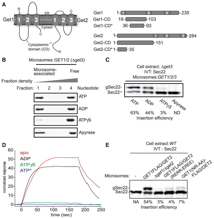
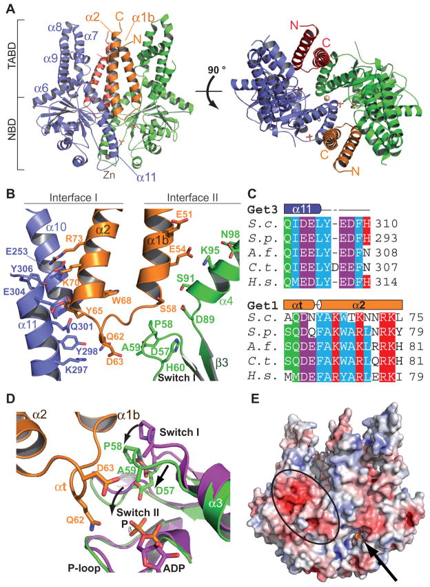
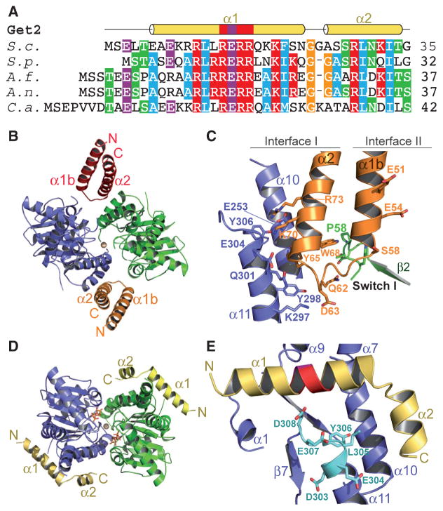
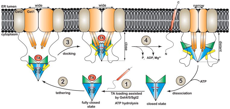
Tail-anchored (TA) proteins are involved in cellular processes including trafficking, degradation, and apoptosis. They contain a C-terminal membrane anchor and are posttranslationally delivered to the endoplasmic reticulum (ER) membrane by the Get3 adenosine triphosphatase interacting with the hetero-oligomeric Get1/2 receptor. We have determined crystal structures of Get3 in complex with the cytosolic domains of Get1 and Get2 in different functional states at 3.0, 3.2, and 4.6 angstrom resolution. The structural data, together with biochemical experiments, show that Get1 and Get2 use adjacent, partially overlapping binding sites and that both can bind simultaneously to Get3. Docking to the Get1/2 complex allows for conformational changes in Get3 that are required for TA protein insertion. These data suggest a molecular mechanism for nucleotide-regulated delivery of TA proteins.


Figures

Fig. 1
Fig. 1
TA protein delivery to the ER membrane by nucleotide-dependent interaction of Get3 with the Get1/2 receptor. (A) Topology models of Get1 and Get2 and an overview of receptor constructs used in this study. Truncated versions of the cytosolic domains (CDs) are marked by an asterisk. (B) Flotation assays of FLAG-tagged Get3 in the presence of different nucleotides or in the apo form with a density gradient and Get1/2/Δ3 microsomes. Nonhydrolyzable ATP-γ-S inhibits targeting of Get3 to the membrane receptor and results in free Get3 at the bottom of the gradient (fraction 4). (C) Membrane insertion assays with Sec22 using Get1/2/3 microsomes to analyze the nucleotide dependence. In vitro translation (IVT) of opsin-tagged Sec22 was performed in the presence of 35S-labeled methionine. Successful insertion is indicated by cargo glycosylation (gSec22). (D) SPR studies on the interaction of Get3 with Get1-CD in the presence of different nucleotides. ATP* indicates measurements with the hydrolysis-deficient D57N mutant and therefore correspond to ATP-γ-S. (E) Analysis of Get1/2 mutants in membrane insertion. Assays were performed as described in Fig. 1C. A Get1/Get2-deletion mutant is not insertion competent (Δget1/Δget2). Mutations in Get2 (14-RERR to EREE) and in Get1 (72-NR to AA) completely abolish insertion, which highlights their equal importance.
Fig. 2
Fig. 2
Structure of the open Get3/Get1 complex. (A) Overall structure shown in views along (left, with indicated domains) and from the membrane (right). Get3 is depicted in blue and green, Get1 in orange and red, respectively. Phosphate ions are drawn as sticks. (B) Close-up of the Get3/Get1 interface. Side chains of residues important for the interaction are shown. The major binding site (interface I) includes helix α2 of Get1 and helices α10 and α11 of Get3 (blue subunit). The DELYED motif (residues 303 to 308) is located at the C terminus of helix α11. The tip of the Get1 coiled-coil contacts the switch I region of the second Get3 subunit (green, interface II). (C) Multiple sequence alignments of the Get3 DELYED motif (top) and the conserved tip region of Get1 (bottom). The respective secondary structures are given on top of the alignments. Color coding is according to conserved charge or hydrophobicity. For species see fig. S1. (D) Get1 is an ATPase effector. Superposition of the Get3 active site within the Get3/Get1 complex (colored as above) with the ADP bound Get3 structure (purple, PDB code 3A37). Get1 pushes the switch regions into the active site as indicated by arrows. Glutamine Q62 within the tip helix (αt) directly binds to the P-loop. (E) Surface potential of the Get3/Get1 complex. Positive charges are indicated in blue, negative charges in red. The view is the same as in Fig. 2A (left). The tunnel leading to the active site of Get3 (shown with modeled ADP) is indicated by an arrow. The prominent negative surface patch on Get3 involving the DELYED motif is marked by an oval.
Fig. 3
Fig. 3
Structure of the semiopen Get3/Get1 and the closed Get3/Get2 complexes. (A) Multiple sequence alignment of Get2 (for species, see fig. S7). The secondary structure is given above the sequences with the RERR motif highlighted. The two helices are connected by a flexible glycine linker. (B) Membrane view of the semiopen Get3/Get1 complex. The complex represents the state halfway between the closed and open Get3 structures. (C) Close-up as shown in Fig. 2B. Although interface I is identical to the open Get3/Get1 complex, interface II changed completely, and Get1 does not contact the switch I region, which is unperturbed compared with the ADP structure shown in Fig. 2D. (D) Membrane view of the closed Get3/Get2 complex with bound ADP-Mg2+ shown in sticks. Get2 (yellow) forms two helices (α1, α2) binding at the periphery of Get3 and contacting only one Get3 subunit. (E) Close-up of the Get3/Get2 interface. Get2 wraps around the DELYED motif (cyan) of Get3 that forms a binding platform. The conserved RERR motif (same color as in Fig. 3A) in the center of helix α1Get2 recognizes the DELYED motif and, together with the additional positive charges, reads out the negative surface patch of Get3 shown in Fig. 2E.
Fig. 4
Fig. 4
Model for TA protein targeting and insertion by the Get pathway. TA protein binding (step 1) locks Get3 in the fully-closed state as inferred from the transition state structure with ADP·AlF4 (PDB code 2WOJ), and bound ATP is hydrolyzed to ADP·Pi. Get3/TA is tethered to Get2 (step 2) as deduced from the Get3/Get2 structure and Get1 associates with the Get3/TA/Get2 complex, which results in an insertion competent complex (step 3). Release of the TA protein relaxes Get3 into the closed state, which allows for Pi release (step 4). The stored energy of ATP hydrolysis drives Get3 via the semiopen to the open state as observed in the Get3/Get1 structures and ADP-Mg2+ is released. Get3 dissociates from the membrane by ATP binding and adopts the closed conformation (step 5). The transition from closed to open Get3 reduces the distance between the two Get1 protomers, whereas the distance between the zinc ion (black dot) and the membrane increases (distances are indicated by arrows, see also fig. S11).

Comment in

  • It takes two to Get3.
    Sinning I, Bange G, Wild K. Sinning I, et al. Structure. 2011 Oct 12;19(10):1353-5. doi: 10.1016/j.str.2011.10.001. Structure. 2011. PMID: 22000508

References

    1. Blobel G, Dobberstein B. J Cell Biol. 1975;67:835. - PMC - PubMed
    1. Schuldiner M, et al. Cell. 2008;134:634. - PMC - PubMed
    1. Borgese N, Brambillasca S, Colombo S. Curr Opin Cell Biol. 2007;19:368. - PubMed
    1. Wattenberg B, Lithgow T. Traffic. 2001;2:66. - PubMed
    1. High S, Abell BM. Biochem Soc Trans. 2004;32:659. - PubMed

Publication types

MeSH terms

Substances