. 2011 Jul 1;333(6038):58-62.
doi: 10.1126/science.1200758.

Probing individual environmental bacteria for viruses by using microfluidic digital PCR

Affiliations

Probing individual environmental bacteria for viruses by using microfluidic digital PCR

Arbel D Tadmor et al. Science. .

Abstract

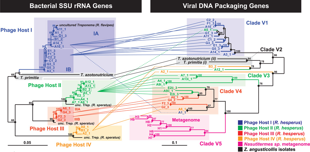
Viruses may very well be the most abundant biological entities on the planet. Yet neither metagenomic studies nor classical phage isolation techniques have shed much light on the identity of the hosts of most viruses. We used a microfluidic digital polymerase chain reaction (PCR) approach to physically link single bacterial cells harvested from a natural environment with a viral marker gene. When we implemented this technique on the microbial community residing in the termite hindgut, we found genus-wide infection patterns displaying remarkable intragenus selectivity. Viral marker allelic diversity revealed restricted mixing of alleles between hosts, indicating limited lateral gene transfer of these alleles despite host proximity. Our approach does not require culturing hosts or viruses and provides a method for examining virus-bacterium interactions in many environments.


Figures

Fig. 1
Fig. 1
End-point fluorescence measured in a panel of a microfluidic digital PCR array. (A) The measured end-point fluorescence from the rRNA channel (right half of each chamber, with the left half masked) and the terminase channel (left half of each chamber, with the right half masked) in a microfluidic array panel. Each panel in the array (1 of 12) consists of 765 reaction chambers 150 µm by 150 µm by 270 µm (6 nl). Retrieved colocalizations are outlined in orange, and positive rRNA chambers randomly selected for retrieval are outlined in gray. FA indicates false alarm (a probable terminase primer-dimer). (B) Normalized amplification curves of all chambers in (A) after linear derivative baseline correction (red, viral; green, rRNA). (C) Specific physical associations between a bacterial cell and the viral marker gene resulting in colocalization include, for example, an attached or assembling virion, injected DNA, an integrated prophage, or a plasmid containing the viral marker gene.
Fig. 2
Fig. 2
Phylogenetic relationship between cultured and uncultured bacterial host rRNA genes and their associated viral DNA packaging genes. (Left) Maximum likelihood (ML) tree of 898 unambiguous nucleotides of the SSU rRNA gene of ribotypes that repeatedly colocalized with the terminase gene, including the two isolated spirochetes Treponema primitia and Treponema azotonutricium. Shorter sequences (A7, 780 bp, and A9, 806 bp) were added by parsimony (dashed branches). (Right) ML tree of 705 unambiguous nucleotides of the large terminase subunit gene. Connecting lines represent colocalized pairs, revealing restricted mixing of terminase alleles between different bacterial hosts. For association of three additional recombinant sequences (boxed on the left), see fig. S5. Statistically, we estimate that an average of 0.6 colocalizations are false [~2% error (19)]. The sequence error rates (40) for the rRNA and terminase genes were measured to be 0 (n = 8) and <0.6 ± 0.3% SD (n = 9), respectively (18). Alleles are named by array (A to G) and retrieval index followed by an underscore and the colony number (colony 1 being sampled 6 months before colonies 2 and 3). Lowercase roman numerals indicate multiple terminases per chromosome. Scale bars represent substitutions per alignment. For interpretation of node support, refer to (18), and for accession numbers, table S10.
Fig. 3
Fig. 3
Rank abundance curve of free-living Treponema spirochetes in R. hesperus termites identifying putative phage hosts. A library of 118 random chambers positive for the rRNA gene were retrieved, postamplified, and sequenced. Of these, n = 78 were related to the Treponema genus, corresponding to 28 different phylotypes based on an operational taxonomical unit, OTU, cut-off set by DOTUR (41) at 3.1%. We show these 28 phylotypes, designated as REPs, ordered by their abundance. Phylotype abundance is expected to reflect true relative abundances in the gut because single-cell amplification is not susceptible to primer bias or rRNA copy number bias. Phylotypes identified as phage hosts are marked by red bars (with the highly clonal marker associated with host I depicted by green viruses and the divergent marker associated with host II depicted by colored viruses). The most abundant free-living Treponema in the gut—REPs 1, 2, and 3 (blue bars)—were not associated with the viral marker. Remaining bars are gray. Error bars are estimated by the binomial SD. See table S5 for OTU assignment. Note that the isolated spirochetes were not spanned by these REPs (fig. S4).

Comment in

References

    1. Suttle CA. Nat. Rev. Microbiol. 2007;5:801. - PubMed
    1. Sullivan MB, et al. Environ. Microbiol. 2010;12:3035. - PMC - PubMed
    1. Lindell D, et al. Nature. 2007;449:83. - PubMed
    1. Angly FE, et al. PLoS Biol. 2006;4:e368. - PMC - PubMed
    1. Williamson SJ, et al. PLoS ONE. 2008;3:e1456. - PMC - PubMed

Publication types

MeSH terms