Comparative Study
. 2012 Jan-Feb;32(1):10-9.
doi: 10.3747/pdi.2010.00200. Epub 2011 Jun 30.

Activation of p38 mitogen-activated protein kinase promotes peritoneal fibrosis by regulating fibrocytes

Affiliations
Comparative Study

Activation of p38 mitogen-activated protein kinase promotes peritoneal fibrosis by regulating fibrocytes

Satoshi Kokubo et al. Perit Dial Int. 2012 Jan-Feb.

Abstract

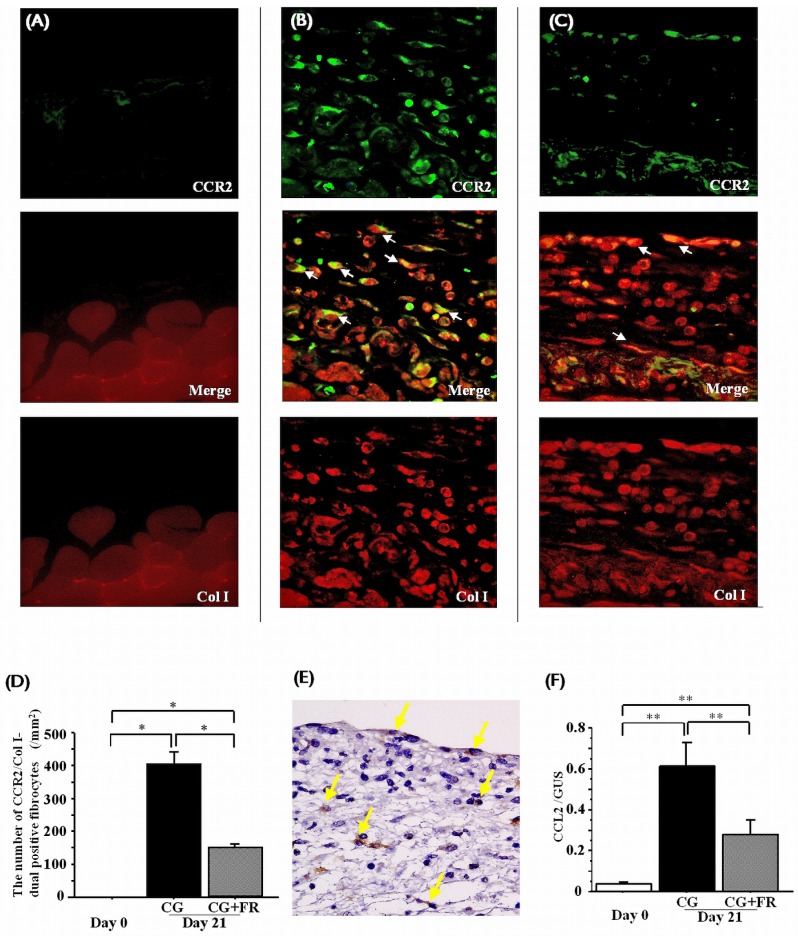
Background: Peritoneal fibrosis is a serious complication of long-term peritoneal dialysis, and yet the precise pathogenic mechanisms of peritoneal fibrosis remain unknown. Fibrocytes participate in tissue fibrosis and express chemokine receptors that are necessary for migration. The p38 mitogen-activated protein kinase (MAPK) pathway regulates the production of chemokines and has been demonstrated to contribute to the pathogenesis of various fibrotic conditions. Accordingly, we used an experimental mouse model of peritoneal fibrosis to examine the dependency of fibrocytes on p38MAPK signaling.

Methods: Peritoneal fibrosis was induced in mice by the injection of 0.1% chlorhexidine gluconate (CG) into the abdominal cavity. Mice were treated with FR167653, a specific inhibitor of p38MAPK, and immunohistochemical studies were performed to detect fibrocytes and cells positive for phosphorylated p38MAPK. The involvement of p38MAPK in the activation of fibrocytes also was also investigated in vitro.

Results: Fibrocytes infiltrated peritoneum in response to CG, and that response was accompanied by progressive peritoneal fibrosis. The phosphorylation of p38MAPK, as defined by CD45+ spindle-shaped cells, was detected both in peritoneal mesothelial cells and in fibrocytes. The level of peritoneal expression of CCL2, a chemoattractant for fibrocytes, was upregulated by CG injection, and treatment with FR167653 reduced the number of cells positive for phosphorylated p38MAPK, the peritoneal expression of CCL2, and the extent of peritoneal fibrosis. Pretreatment with FR167653 inhibited the expression of procollagen type I α1 induced by transforming growth factor-β1.

Conclusions: Our results suggest that p38MAPK signaling contributes to peritoneal fibrosis by regulating fibrocyte function.


Figures

Figure 1
Figure 1
— Peritoneal thickness and type I collagen α1 (COLIAI) expression in the murine peritoneum. (A) Normal control at day 0 shows no evidence of peritoneal thickness (200× original magnification). (B) Peritoneal thickness at 21 days after the start of chlorhexidine gluconate (CG) injections (“CG mice”) (200× original magnification). (C) Peritoneal thickness at day 21 in mice treated with FR167653 (“CG+FR mice”) (200× original magnification). (D) Injection with CG caused the peritoneum to thicken in CG mice. By contrast, the peritoneum was less thick in CG+FR mice. (E) Upregulation of COLIAI messenger RNA expression was induced by CG injection and reduced with FR167653 treatment. Values are mean ± standard error of the mean. * p < 0.01. ** p < 0.05.
Figure 2
Figure 2
— Fibrocyte infiltration into peritoneum. Dual immunostaining for CD45 and type I collagen (Col I) was performed (green = CD45; yellow = dual-positive fibrocyte (arrows); red = Col I). Dual-positive fibrocytes infiltrated peritoneum after chlorhexidine gluconate (CG) injection. (A) Normal control at day 0 (200× original magnification). (B) Mice injected with CG (200× original magnification). (C) Mice injected with CG and treated with FR167653 (CG+FR) (200× original magnification). Arrows indicate cells that are dually positive for CD45 and Col I. (D) The number of infiltrating fibrocytes was lower in mice treated with FR167653. Values are mean ± standard error of the mean. * p < 0.01.
Figure 3
Figure 3
— Fibrocytes expressing CCR2, and CCL2 production, in peritoneum. Dual immunostaining for CCR2 and type I collagen (Col I) was performed (green = CCR2; yellow = CCR2-positive fibrocytes (arrows); red = Col I). Fibrocytes positive for CCR2 were detected in peritoneum after chlorhexidine gluconate (CG) injection. (A) Normal control at day 0. (B) Mice injected with CG. White arrows indicate cells positive for both CCR2 and Col I (200× original magnification). (C) Mice injected with CG and treated with FR167653 (CG+FR). White arrows indicate cells positive for both CCR2 and Col I (200× original magnification). (D) Fewer infiltrating CCR2-positive fibrocytes were observed in CG+FR mice. (E) Production of the CCL2 protein was induced by CG injection in peritoneal mesothelial cells as well as in interstitial cells. Yellow arrows indicate CCL2-positive cells (200× original magnification). (F) After CG injection, messenger RNA transcripts for CCL2 were upregulated in peritoneum; in CG+FR mice, levels of CCL2 were lower. Values are mean ± standard error of the mean. * p < 0.01. ** p < 0.05.
Figure 4
Figure 4
— The presence of cells positive for phosphorylated p38 mitogen-activated protein kinase (p-p38MAPK) in peritoneum. (A) Arrowheads indicate p-p38MAPK–positive mesothelial cells. Arrows indicate p-p38MAPK–positive spindle-shaped cells (200× original magnification). (B) The percentage of p-p38MAPK–positive mesothelial cells in peritoneum was lower in mice treated with FR167653 (CG+FR). (C) The number of p38MAPK-positive interstitial cells in peritoneum was significantly reduced in CG+FR mice. Values are mean ± standard error of the mean. * p < 0.01.
Figure 5
Figure 5
— Characterization of peritoneal cells positive for phosphorylated p38 mitogen-activated protein kinase (p-p38MAPK). Dual immunostaining for CD45 and p-p38MAPK was performed (brown = p-p38MAPK; red = CD45). Arrows indicate spindle-shaped cells positive for CD45 and p-p38MAPK. (A) Normal control at day 0 (200× original magnification). (B) Mice injected with CG (200× original magnification). (C) Mice injected with CG and treated with FR167653 (CG+FR) (200× original magnification). (D) The number of spindle-shaped cells positive for CD45 and p-p38MAPK was lower in CG+FR mice. Values are mean ± standard error of the mean. * p < 0.01. ** p < 0.05.
Figure 6
Figure 6
— The effect of p38 mitogen-activated protein kinase on production of procollagen type I α1 (COLIAI). Stimulation with transforming growth factor β1 (TGF) induced isolated fibrocytes to produce COLIAI messenger RNA, an effect that was reduced by treatment with FR167653 (FR). Values are mean ± standard error of the mean. * p < 0.05. GAPDH = glyceraldehyde 3-phosphate dehydrogenase.
Figure 7
Figure 7
— Proposed schema for p38 mitogen-activated protein kinase (p38MAPK) signaling in the pathogenesis of peritoneal fibrosis: regulation of fibrocyte infiltration by production of CCL2 (a chemoattractant for fibrocytes), and production of collagen in fibrocytes.

Comment in

Similar articles

Cited by

References

    1. Selgas R, Fernandez–Reyes MJ, Bosque E, Bajo MA, Borrego F, Jimenez C, et al. Functional longevity of the human peritoneum: how long is continuous peritoneal dialysis possible? Results of a prospective medium long-term study. Am J Kidney Dis 1994; 23:64–73 - PubMed
    1. Davies SJ, Bryan J, Phillips L, Russell GI. Longitudinal changes in peritoneal kinetics: the effects of peritoneal dialysis and peritonitis. Nephrol Dial Transplant 1996; 11:498–506 - PubMed
    1. Cnossen TT, Konings CJ, Kooman JP, Lindholm B. Peritoneal sclerosis—aetiology, diagnosis, treatment and prevention. Nephrol Dial Transplant 2006; 21(Suppl 2):ii38–41 - PubMed
    1. Phillips RJ, Burdick MD, Hong K, Lutz MA, Murray LA, Xue YY, et al. Circulating fibrocytes traffic to the lungs in response to CXCL12 and mediate fibrosis. J Clin Invest 2004; 114:438–46 - PMC - PubMed
    1. Sakai N, Wada T, Yokoyama H, Lipp M, Ueha S, Matsushima K, et al. Secondary lymphoid tissue chemokine (SLC/CCL21)/CCR7 signaling regulates fibrocytes in renal fibrosis. Proc Natl Acad Sci U S A 2006; 103:14098–103 - PMC - PubMed

Publication types

MeSH terms