. 2011 Jun;23(6):2125-42.
doi: 10.1105/tpc.111.085340. Epub 2011 Jun 30.

STENOFOLIA regulates blade outgrowth and leaf vascular patterning in Medicago truncatula and Nicotiana sylvestris

Affiliations

STENOFOLIA regulates blade outgrowth and leaf vascular patterning in Medicago truncatula and Nicotiana sylvestris

Million Tadege et al. Plant Cell. 2011 Jun.

Abstract

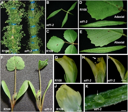
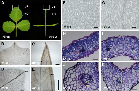
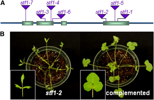
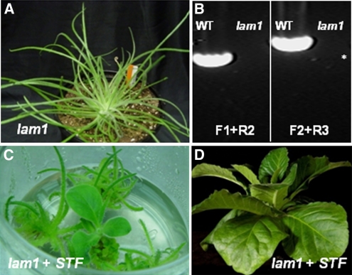
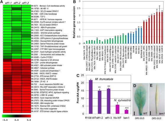
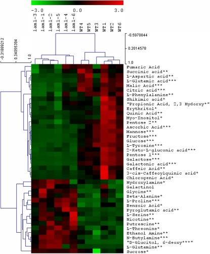
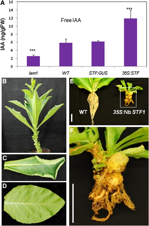
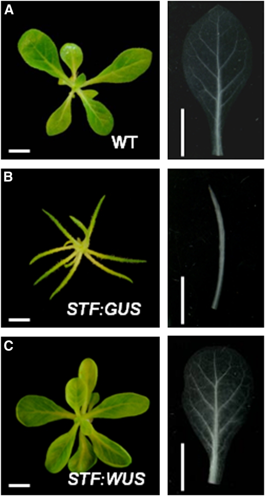
Dicot leaf primordia initiate at the flanks of the shoot apical meristem and extend laterally by cell division and cell expansion to form the flat lamina, but the molecular mechanism of lamina outgrowth remains unclear. Here, we report the identification of STENOFOLIA (STF), a WUSCHEL-like homeobox transcriptional regulator, in Medicago truncatula, which is required for blade outgrowth and leaf vascular patterning. STF belongs to the MAEWEST clade and its inactivation by the transposable element of Nicotiana tabacum cell type1 (Tnt1) retrotransposon insertion leads to abortion of blade expansion in the mediolateral axis and disruption of vein patterning. We also show that the classical lam1 mutant of Nicotiana sylvestris, which is blocked in lamina formation and stem elongation, is caused by deletion of the STF ortholog. STF is expressed at the adaxial-abaxial boundary layer of leaf primordia and governs organization and outgrowth of lamina, conferring morphogenetic competence. STF does not affect formation of lateral leaflets but is critical to their ability to generate a leaf blade. Our data suggest that STF functions by modulating phytohormone homeostasis and crosstalk directly linked to sugar metabolism, highlighting the importance of coordinating metabolic and developmental signals for leaf elaboration.


Figures

Figure 1.
Figure 1.
Morphological Phenotypes of stf Mutants. (A) Adult M. truncatula genotype R108 (wild type) and stf1-2 mutant plant 9 weeks after transfer to soil. (B) stf1-2 adult leaf showing trifoliate identity and normal proximodistal growth but drastically affected mediolateral growth. (C) R108 adult leaf. (D) Adaxial surface of R108 and stf adult leaves where margin serrations are absent in the mutant. (E) Abaxial surface of R108 and stf adult leaves where major veins are not visible in the mutant. (F) R108 and stf seedlings at the unifoliate (first leaf) stage where cotyledons are nearly wild type and the unifoliate leaf is partially affected. (G) R108 flower before anthesis in which the anthers and stigma are enclosed by petal. (H) stf flower in the same stage as in (G) but with anthers and stigma exposed (arrow) because of the narrow petal. (I) stf outer petal showing the reduction in lateral expansion. (J) R108 outer petal in the same stage as in (I). (K) stf ovary wall failing to close and ovule protruding out (arrow). Scale bars in (I) and (J) = 1 mm and in (K) = 50 μM.
Figure 2.
Figure 2.
The stf Mutant Is Severely Defective in Leaf Vascular Patterning. (A) Wild-type, R108, and stf1-2 mature leaves showing the regions for close-up described in (B) to (E). (B) to (E) Leaf material observed through a light microscope after clearing with lactic acid. (B) Major and minor veins of R108 leaf. (C) Disorganized and poorly developed major veins in stf. Major veins are forming near the margins (one on either side of the midvein) along the proximodistal axis (arrows). (D) R108 major vein extends close to the margin with its tip aligned to the serration and is open ended (arrow). (E) stf major vein poorly developed and connected to marginal vein (arrow). (F) R108 leaf epidermal cells viewed through a light microscope. (G) Epidermal cells of stf leaf showing narrower width. (H) Transverse section through R108 leaf blade showing palisade mesophyll (white arrow) and spongy mesophyll (red arrow) cells. Sections were stained with Toluidine Blue. (I) Transverse section through stf leaf blade showing the poor distinction between palisade mesophyll (white arrow) and spongy mesophyll (red arrow) cells. (J) Transverse section through the midrib of R108 leaf showing xylem (yellow arrow) and phloem (orange arrow) vessels. (K) Transverse section through stf midrib showing poorly differentiated xylem and phloem vessels (yellow and orange arrows) and cortical tissue. Scale bars in (B) to (E) = 500 μM, in (F) and (G) = 50 μM, and in (H) to (K) = 100 μM.
Figure 3.
Figure 3.
STF Encodes a WOX Domain Protein and Complements the stf Mutant. (A) STF gene structure showing the position of the Tnt1 insertion site in seven independent mutant lines. (B) stf mutant complemented with 5.3-kb genomic STF. [See online article for color version of this figure.]
Figure 4.
Figure 4.
STF Expression Pattern in Vegetative and Floral Apices by RNA in Situ Hybridization in R108. (A) STF expression in 12-d-old vegetative shoot apex viewed in longitudinal sections. At very early stages, STF is adaxially expressed in few cells (black arrows), but absent from the central zone of SAM (white arrow). (B) STF expression in older leaf primordia showing localization at the adaxial–abaxial boundary layer (arrow). (C) STF expression in young flower showing a strong signal in petal primordia and developing petal (red arrows) and developing carpel (black arrow). (D) STF expression in mature flower showing strong localization in the placenta at the base of the ovules (arrow). (E) STF expression in mature flower showing expression in the petal lobe (arrow). (F) STF expression in inflorescence apex showing activity in floral organ primordia, but no detection in the inflorescence meristem (white arrow). (G) STF expression in inflorescence apex showing no detection in the floral meristem (white arrow). (H) PIM (AP1) expression in inflorescence apex shown here as positive control for expression in floral meristem (arrow). (I) RNA in situ hybridization in the inflorescence apex using STF sense probe as negative control.
Figure 5.
Figure 5.
The N. sylvestris lam1 Mutation Is Caused by Deletion of the Ns STF1 Gene. (A) Ten-week-old adult lam1 mutant plant. (B) Genomic PCR showing deletion of the Ns STF1 locus in the lam1 mutant. Primers F1+R2 amplify the complete CDS plus the 3′ UTR, and primers F2+R3 amplify the promoter, the 5′ UTR, plus part of the CDS, and together span 5.67 kb of the Ns STF1 region. * = 3 kb. WT, wild type. (C) Untransformed lam1 mutant and lam1 complemented with M. truncatula 5.3-kb genomic STF regenerating in nonselective tissue culture media. (D) Complemented lam1 in (C) 4 weeks after transfer to soil.
Figure 6.
Figure 6.
Microarray Analysis and Auxin Quantification in stf and lam1 Mutants. (A) A heat map showing differentially expressed genes in three stf mutant lines compared with wild type R108 in 4-week-old shoot apices. Representative genes that are downregulated (green) and upregulated (red) with twofold or more difference are shown. ABA, abscisic acid. (B) Validation of relative gene expression of selected genes in the stf mutant compared with the wild type by qRT-PCR. Wild-type expression level was arbitrarily set to 1.0. Green, downregulated genes; red, upregulated genes; blue, genes not detected by the microarray. Values are the mean and se of three biological replicates. (C) Free IAA content in 4-week-old leaves of stf and lam1 mutants compared with their wild type (WT). Values are the mean and se of five experiments (***P < 0.001, **P < 0.01). (D) GUS staining in DR5:GUS-transformed wild-type N. sylvestris leaf. (E) GUS staining in DR5:GUS-transformed lam1 leaf showing reduced auxin.
Figure 7.
Figure 7.
Metabolic Profiling in 4-Week-Old Leaves of Wild-Type and lam1 Mutant N. sylvestris. Representative common metabolites are shown. Colors indicate downregulated (green) and upregulated (red) metabolites in lam1 mutant compared with the wild type (WT). The black color shows metabolites that are unchanged. The numbers 1 through 6 at the top indicate replicates of lam1 and wild-type samples each from six individual plants. Statistical significance was calculated using Student’s t test (***P < 0.001, **P < 0.01, and *P < 0.05).
Figure 8.
Figure 8.
Ectopic Expression of Mt STF and Nb STF1 in N. sylvestris. (A) Free IAA content in mature leaves of lam1 mutant, Wild type, STF:GUS transgenic control, and 35S:STF transgenic plant. Values are the mean and se of five replicates (***P < 0.001). FW, fresh weight. (B) 35S:STF transgenic plant showing upward curling leaf phenotype. (C) 35S:Nb-STF1 transgenic leaf showing upward curling phenotype. (D) Wild-type leaf. (E) Left, 4-week-old wild-type (WT) N. sylvestris; right, 17-month-old transgenic plant with highest STF overexpression showing shoot and root deformation. (F) Close-up of the transgenic plant in (E) showing two large tumors. Scale bars = 5 cm.
Figure 9.
Figure 9.
Exogenous Application of Auxin and Cytokinin Partially Rescues the lam1 Lamina. (A) and (B) Wild-type N. sylvestris (A) and lam1 (B) shoots treated with 10 mM BAP. Inset shows leaf branching. (C) and (D) Wild-type N. sylvestris (C) and lam1 (D) shoots treated with 10 mM BAP plus 10 mM IAA. Inset shows partially formed petiole and blade. (E) and (F) Wild-type N. sylvestris (E) and lam1 (F) shoots treated with 10 mM BAP plus 1 mM IAA. Inset shows partially formed petiole and blade. Note that lam1 leaves are uniformly thin and cannot be distinguished into petiole and lamina. Scale bars = 1 cm. [See online article for color version of this figure.]
Figure 10.
Figure 10.
Arabidopsis WUS Complements the lam1 Mutant Phenotype. (A) Untransformed wild-type (WT) N. sylvestris grown in tissue culture MS media. Right panel shows wild-type leaf cleared with lactic acid for looking at the venation pattern. (B) lam1 mutant transformed with STF:GUS construct as negative control. Right panel shows lam1 leaf cleared with lactic acid. (C) lam1 mutant complemented with STF:WUS construct. Right panel shows complemented leaf cleared with lactic acid. Note that the lamina and venation phenotypes are complemented. [See online article for color version of this figure.]

Similar articles

Cited by

References

    1. Aida M., Ishida T., Fukaki H., Fujisawa H., Tasaka M. (1997). Genes involved in organ separation in Arabidopsis: An analysis of the cup-shaped cotyledon mutant. Plant Cell 9: 841–857 - PMC - PubMed
    1. Ariel F., Diet A., Verdenaud M., Gruber V., Frugier F., Chan R., Crespi M. (2010). Environmental regulation of lateral root emergence in Medicago truncatula requires the HD-Zip I transcription factor HB1. Plant Cell 22: 2171–2183 - PMC - PubMed
    1. Barkoulas M., Hay A., Kougioumoutzi E., Tsiantis M. (2008). A developmental framework for dissected leaf formation in the Arabidopsis relative Cardamine hirsuta. Nat. Genet. 40: 1136–1141 - PubMed
    1. Benková E., Michniewicz M., Sauer M., Teichmann T., Seifertová D., Jürgens G., Friml J. (2003). Local, efflux-dependent auxin gradients as a common module for plant organ formation. Cell 115: 591–602 - PubMed
    1. Bilsborough G.D., Runions A., Barkoulas M., Jenkins H.W., Hasson A., Galinha C., Laufs P., Hay A., Prusinkiewicz P., Tsiantis M. (2011). Model for the regulation of Arabidopsis thaliana leaf margin development. Proc. Natl. Acad. Sci. USA 108: 3424–3429 - PMC - PubMed

Publication types

MeSH terms