. 2011 Aug 26;286(34):29511-20.
doi: 10.1074/jbc.M111.236810. Epub 2011 Jun 30.

Identification of a negative regulatory region for the exchange activity and characterization of T332I mutant of Rho guanine nucleotide exchange factor 10 (ARHGEF10)

Affiliations

Identification of a negative regulatory region for the exchange activity and characterization of T332I mutant of Rho guanine nucleotide exchange factor 10 (ARHGEF10)

Taro Chaya et al. J Biol Chem. .

Abstract

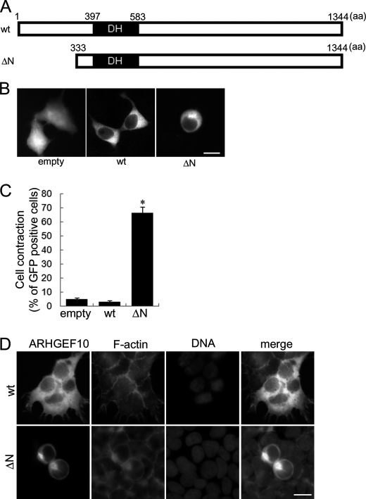
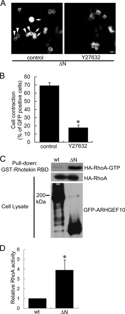
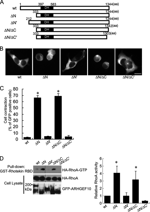
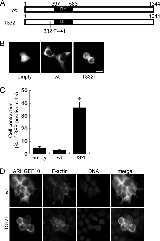
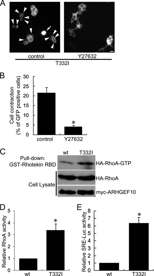
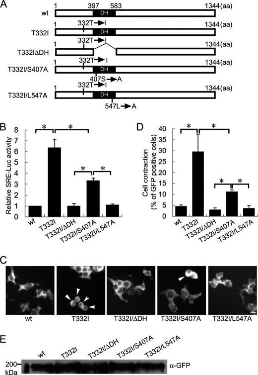
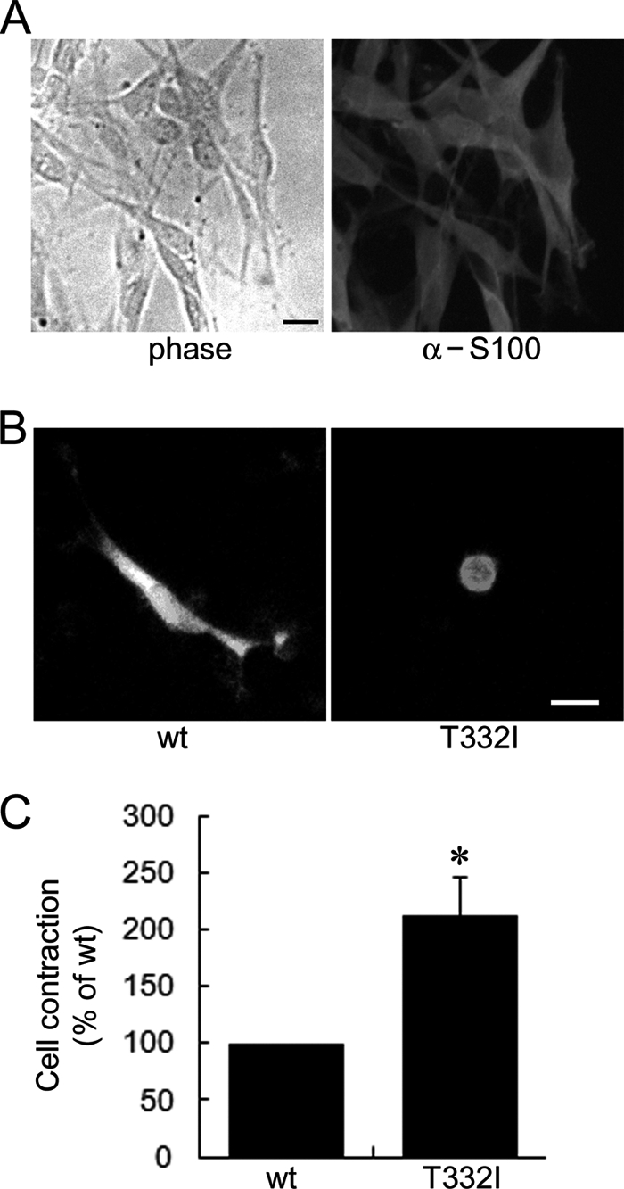
The T332I mutation in Rho guanine nucleotide exchange factor 10 (ARHGEF10) was previously found in persons with slowed nerve conduction velocities and thin myelination of peripheral nerves. However, the molecular and cellular basis of the T332I mutant is not understood. Here, we show that ARHGEF10 has a negative regulatory region in the N terminus, in which residue 332 is located, and the T332I mutant is constitutively active. An N-terminal truncated ARHGEF10 mutant, ARHGEF10 ΔN (lacking amino acids 1-332), induced cell contraction that was inhibited by a Rho kinase inhibitor Y27632 and had higher GEF activity for RhoA than the wild type. The T332I mutant also showed the phenotype similar to the N-terminal truncated mutant. These data suggest that the ARHGEF10 T332I mutation-associated phenotype observed in the peripheral nerves is due to activated GEF activity of the ARHGEF10 T332I mutant.


Figures

FIGURE 1.
FIGURE 1.
Cell contraction induced by ARHGEF10 ΔN in HEK293T cells. A, domain structures of ARHGEF10 are presented. Full-length ARHGEF10 (wt) and an N-terminal truncation mutant lacking N-terminal 332 amino acids of ARHGEF10 (ΔN) are shown. B, HEK293T cells were transiently transfected with the plasmid encoding GFP, GFP-tagged ARHGEF10 wt, or GFP-ARHGEF10 ΔN. After 48 h, fluorescence images of living cells were observed. Scale bar, 10 μm. C, quantitative analysis of cell contraction of HEK293T cells is shown. HEK293T cells were transiently transfected with the plasmid encoding GFP, GFP-ARHGEF10 wt or ΔN. The proportion of cell contraction was scored as a percentage of the rounded cells of GFP-positive cells. Cells floating in the culture medium were excluded from counting. The data represent the mean ± S.E. (error bars) from three independent experiments. *, p < 0.05 compared with empty. For each experiment, >100 cells were counted. D, HEK293T cells were transiently transfected with the plasmid encoding GFP-ARHGEF10 wt or ΔN. After 48 h, cells were fixed and co-stained with TRITC-phalloidin (F-actin) and Hoechst33258 (DNA). Transfected cells are shown by the fluorescence of GFP. Scale bar, 10 μm.
FIGURE 2.
FIGURE 2.
ARHGEF10 ΔN-induced cell contraction mediated through Rho-ROCK signaling. A, HEK293T cells were transiently transfected with the plasmid encoding GFP-ARHGEF10 ΔN and treated with or without 10 μm Y27632 for 24 h. After that, fluorescence images of living cells were observed. Arrowheads indicate rounded cells. Arrows indicate cells floating in the culture medium. Scale bar, 10 μm. B, quantitative analysis of cell contraction of HEK293T cells. HEK293T cells were transiently transfected with the plasmid encoding GFP-ARHGEF10 ΔN and treated with or without 10 μm Y27632 for 24 h. The proportion of cell contraction was scored as a percentage of the rounded cells of GFP-positive cells. Cells floating in the culture medium were excluded from counting. The data represent the mean ± S.E. (error bars) from three independent experiments. *, p < 0.05. For each experiment, >100 cells were counted. C and D, HEK293T cells were transiently co-transfected with the plasmids encoding GFP-ARHGEF10 wt or ΔN, and HA-tagged RhoA. After 48 h, cells were lysed with lysis buffer. The supernatants were mixed with GST-fused Rhotekin-RBD, and an active form of RhoA (RhoA-GTP) was pulled down. The amounts of active form of RhoA were determined by immunoblotting with anti-HA antibody. Expressions of HA-RhoA and GFP-ARHGEF10 in cell lysates were also detected by immunoblotting with anti-HA and anti-GFP antibodies, respectively. The representative image of three independent experiments is shown. Relative RhoA activity is indicated by the amounts of RhoA-GTP normalized to total amounts of RhoA in cell lysates, and values are expressed as -fold value of cells transfected with wt. The data are the mean ± S.E. from three independent experiments. *, p < 0.05.
FIGURE 3.
FIGURE 3.
Cell contraction and activation of RhoA by various deletion mutants of ARHGEF10 in HEK293T cells. A, domain structures of ARHGEF10. Full-length ARHGEF10 (wt), N-terminal truncation mutants lacking N-terminal 332 and 211 amino acids of ARHGEF10 (ΔN and ΔN′, respectively), and C-terminal truncation mutants lacking C-terminal 2 and 41 amino acids of ARHGEF10 ΔN (ΔN/ΔC and ΔN/ΔC′, respectively) are shown. B, HEK293T cells transiently transfected with the plasmid encoding GFP-ARHGEF10 wt, ΔN, ΔN′, ΔN/ΔC, or ΔN/ΔC′. After 48 h, fluorescence images of living cells observed. Scale bar, 10 μm. C, quantitative analyses of cell contraction of HEK293T cells. HEK293T cells were transiently transfected with the plasmid encoding GFP-ARHGEF10 wt, ΔN, ΔN′, ΔN/ΔC, or ΔN/ΔC′. The proportion of cell contraction was scored as a percentage of the rounded cells of GFP-positive cells. Cells floating in the culture medium were excluded from counting. The data represent the mean ± S.E. (error bars) from three independent experiments. *, p < 0.05 compared with wt. For each experiment, >100 cells were counted. D, HEK293T cells transiently co-transfected with the plasmids encoding GFP-ARHGEF10 wt, ΔN, ΔN′, ΔN/ΔC, or ΔN/ΔC′, and HA-RhoA. After 48 h, cells were lysed with lysis buffer. The supernatants were mixed with GST-Rhotekin-RBD, and an active form of RhoA was pulled down. The amounts of active form of RhoA were determined by immunoblotting with anti-HA antibody. Expressions of HA-RhoA and GFP-ARHGEF10 in cell lysates were also detected by immunoblotting with anti-HA and anti-GFP antibodies, respectively. The representative image of three independent experiments is shown. Relative RhoA activity is indicated by the amounts of RhoA-GTP normalized to total amounts of RhoA in cell lysates, and values are expressed as -fold value of cells transfected with wt. The data are the mean ± S.E. from three independent experiments. *, p < 0.05 compared with wt.
FIGURE 4.
FIGURE 4.
Cell contraction induced by ARHGEF10 T332I in HEK293T cells. A, domain structures of ARHGEF10. Full-length ARHGEF10 (wt) and the T332I mutant of full-length ARHGEF10 are shown. B, HEK293T cells transiently transfected with the plasmid encoding GFP, GFP-ARHGEF10 wt, or GFP-ARHGEF10 T332I. After 48 h, fluorescence images of living cells were observed. Scale bar, 10 μm. C, quantitative analysis of cell contraction of HEK293T cells. HEK293T cells were transiently transfected with the plasmid encoding GFP, GFP-ARHGEF10 wt, or T332I. The proportion of cell contraction was scored as a percentage of the rounded cells of GFP-positive cells. Cells floating in the culture medium were excluded from counting. The data represent the mean ± S.E. (error bars) from three independent experiments. *, p < 0.05 compared with empty. For each experiment, >100 cells were counted. D, HEK293T cells transiently transfected with the plasmid encoding GFP-ARHGEF10 wt or T332I. After 48 h, cells were fixed and co-stained with TRITC-phalloidin (F-actin) and Hoechst33258 (DNA). Transfected cells are shown by the fluorescence of GFP. Scale bar, 10 μm.
FIGURE 5.
FIGURE 5.
ARHGEF10 T332I-induced cell contraction mediated through Rho-ROCK signaling. A, HEK293T cells transiently transfected with the plasmid encoding GFP-ARHGEF10 T332I and treated with or without 10 μm Y27632 for 24 h. Then, fluorescence images of living cells were observed. Arrowheads indicate rounded cells. Arrows indicate cells floating in the culture medium. Scale bar, 10 μm. B, quantitative analysis of cell contraction of HEK293T cells. HEK293T cells were transiently transfected with the plasmid encoding GFP-ARHGEF10 T332I and treated with or without 10 μm Y27632 for 24 h. The proportion of cell contraction was scored as a percentage of the rounded cells of GFP-positive cells. Cells floating in the culture medium were excluded from counting. The data represent the mean ± S.E. (error bars) from three independent experiments. *, p < 0.05. For each experiment, >100 cells were counted. C and D, HEK293T cells transiently co-transfected with the plasmids encoding myc-tagged ARHGEF10 wt or T332I, and HA-RhoA. After 48 h, cells were lysed with lysis buffer. The supernatants were mixed with GST-Rhotekin-RBD, and an active form of RhoA was pulled down. The amounts of active form of RhoA were determined by immunoblotting with anti-HA antibody. Expressions of HA-RhoA and myc-ARHGEF10 in cell lysates were also detected by immunoblotting with anti-HA and anti-myc antibodies, respectively. A representative image of three independent experiments is shown. Relative RhoA activity is indicated by the amounts of RhoA-GTP normalized to total amounts of RhoA in cell lysates, and values are expressed as -fold value of cells transfected with wt. The data are the mean ± S.E. from three independent experiments. *, p < 0.05. E, HEK293T cells transiently co-transfected with the plasmid encoding GFP-ARHGEF10 wt or T332I, pSRE.l-luciferase reporter plasmid encoding firefly luciferase, and pRL-TK control vector encoding Renilla luciferase. The firefly luciferase activities were normalized to the Renilla luciferase activities, and values are expressed as -fold induction compared with wt. The data are the mean ± S.E. from three independent experiments. *, p < 0.05.
FIGURE 6.
FIGURE 6.
Effect of DH mutations of ARHGEF10 T332I on activation of Rho and cell contraction. A, domain structures of ARHGEF10. Full-length ARHGEF10 (wt) and the T332I mutant of full-length ARHGEF10, a DH domain truncation mutant lacking amino acids 397–583 of ARHGEF10 T332I (T332I ΔDH), and the S407A and L547A mutants of ARHGEF10 T332I (T332I/S407A and T332I/L547A, respectively) are shown. B, HEK293T cells transiently co-transfected with the plasmid encoding GFP-ARHGEF10 wt, T332I, T332I ΔDH, T332I/S407A, or T332I/L547A, pSRE.l-luciferase reporter plasmid encoding firefly luciferase, and pRL-TK control vector encoding Renilla luciferase. The firefly luciferase activities were normalized to the Renilla luciferase activities, and values are expressed as -fold induction compared with wt. The data are the mean ± S.E. (error bars) from three independent experiments. *, p < 0.05. C, HEK293T cells transiently transfected with the plasmid encoding GFP-ARHGEF10 wt, T332I, T332I ΔDH, T332I ΔS407A, or T332I/L547A. After 48 h, fluorescence images of living cells were observed. Arrowheads indicate rounded cells. Scale bar, 10 μm. D, quantitative analysis of cell contraction of HEK293T cells. HEK293T cells were transiently transfected with the plasmid encoding GFP-ARHGEF10 wt, T332I, T332I ΔDH, T332I/S407A, or T332I/L547A. The proportion of cell contraction was scored as a percentage of the rounded cells of GFP-positive cells. Cells floating in the culture medium were excluded from counting. The data represent the mean ± S.E. from three independent experiments. *, p < 0.05. For each experiment, >100 cells were counted. E, protein expressions of GFP-ARHGEF10 wt, T332I, T332I ΔDH, T332I/S407A, and T332I/L547A were confirmed by immunoblotting with anti-GFP antibody.
FIGURE 7.
FIGURE 7.
Cell contraction induced by ARHGEF10 T332I in Schwann cells. A, left, phase contrast image of Schwann cells derived from rat sciatic nerves. Right, Schwann cells fixed and stained with anti-S100 antibody. Scale bar, 10 μm. B, Schwann cells transiently transfected with the plasmid encoding GFP-ARHGEF10 wt or T332I. After 48 h, fluorescence images of living cells were observed. Scale bar, 10 μm. C, quantitative analysis of cell contraction of Schwann cells. Schwann cells were transiently transfected with the plasmid encoding GFP-ARHGEF10 wt or T332I. The proportion of cell contraction was scored as a percentage of the rounded cells of GFP-positive cells, and values are expressed as -fold change compared with wt. Cells floating in the culture medium were excluded from counting. The data represent the mean ± S.E. (error bars) from three independent experiments. *, p < 0.05. For each experiment, >30 cells were counted.

Similar articles

Cited by

References

    1. Etienne-Manneville S., Hall A. (2002) Nature 420, 629–635 - PubMed
    1. Burridge K., Wennerberg K. (2004) Cell 116, 167–179 - PubMed
    1. Ridley A. J., Hall A. (1992) Cell 70, 389–399 - PubMed
    1. Ridley A. J., Paterson H. F., Johnston C. L., Diekmann D., Hall A. (1992) Cell 70, 401–410 - PubMed
    1. Nobes C. D., Hall A. (1995) Cell 81, 53–62 - PubMed

Publication types

MeSH terms

LinkOut - more resources