. 2011 Jul;22(7):1262-74.
doi: 10.1681/ASN.2010090970. Epub 2011 Jun 30.

Parietal epithelial cells participate in the formation of sclerotic lesions in focal segmental glomerulosclerosis

Affiliations

Parietal epithelial cells participate in the formation of sclerotic lesions in focal segmental glomerulosclerosis

Bart Smeets et al. J Am Soc Nephrol. 2011 Jul.

Abstract

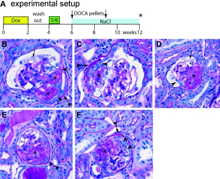
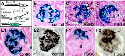
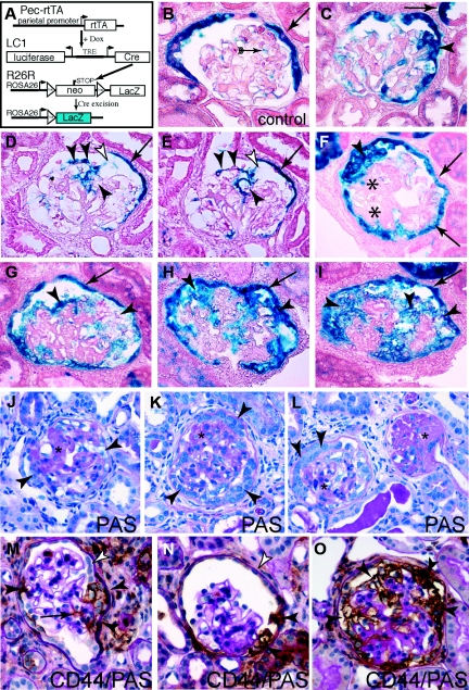
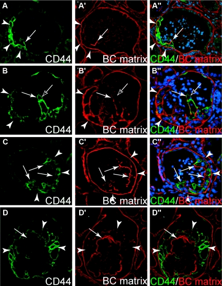
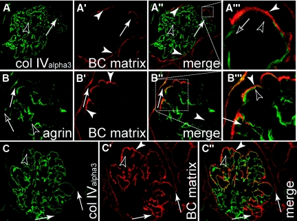
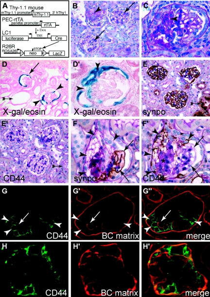
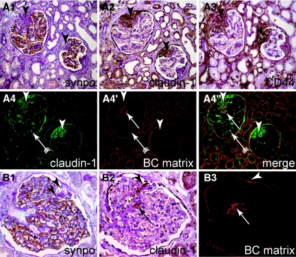
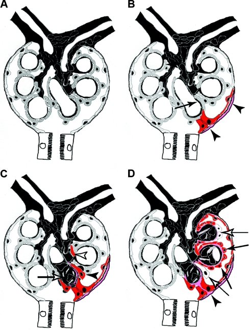
The pathogenesis of the development of sclerotic lesions in focal segmental glomerulosclerosis (FSGS) remains unknown. Here, we selectively tagged podocytes or parietal epithelial cells (PECs) to determine whether PECs contribute to sclerosis. In three distinct models of FSGS (5/6-nephrectomy + DOCA-salt; the murine transgenic chronic Thy1.1 model; or the MWF rat) and in human biopsies, the primary injury to induce FSGS associated with focal activation of PECs and the formation of cellular adhesions to the capillary tuft. From this entry site, activated PECs invaded the affected segment of the glomerular tuft and deposited extracellular matrix. Within the affected segment, podocytes were lost and mesangial sclerosis developed within the endocapillary compartment. In conclusion, these results demonstrate that PECs contribute to the development and progression of the sclerotic lesions that define FSGS, but this pathogenesis may be relevant to all etiologies of glomerulosclerosis.


Figures

Figure 1.
Figure 1.
The 5/6 Nx + DOCA-salt model induces glomerular lesions characteristic of focal and segmental glomerulosclerosis. (A) Schematic of the experimental set-up. After 14 days of treatment with doxycycline (dox) to induce the irreversible genetic labeling and a washout phase of 7 days, a subtotal nephrectomy (5/6) was performed. After a recovery phase, two 25-mg DOCA pellets were implanted subcutaneously (arrows) and 0.1 to 0.9% NaCl was added to the drinking water. Animals were killed after 12 weeks (asterisk). (B through F) PAS-stained paraffin section showing segmental glomerulosclerosis induced in the 5/6 Nx + DOCA-salt model. (B) Lesion near the vascular stalk, where two adhesions (arrowheads), segmental sclerosis (asterisks), a thickened BC (arrow with tails), and vacuolization of podocytes (arrow), is present. (C,D,F) Glomerular lesions with extracapillary proliferations (arrowheads) were also observed. Thickening of the basement membrane of BC (arrow with tails) was observed, mostly in association with an adhesion (B, arrowheads) and/or segmental sclerosis of the glomerular tuft (asterisk). (D and F) More advanced sclerotic lesions (asterisk).
Figure 2.
Figure 2.
Genetically labeled podocytes are absent from sclerotic lesions. (A) Schematic of the podocyte-specific inducible irreversible genetic tagging. For a detailed description see text. (B) Normal β-gal staining of genetically labeled podocytes (blue), as can be observed in control animals after dox administration (control), or in unaffected glomeruli of animals after 5/6 Nx + DOCA-salt treatment. (C and D) In experimental animals, β-gal staining is absent from segmental sclerotic lesions (asterisks) but still preserved within the unaffected portion of the same glomerulus (arrows, X-gal/eosin stained cryosections). (E and F) Serial cryosections were stained for the genetic marker β-gal (E1, F1, blue staining, eosin counterstain) or synaptopodin (E2, F2, brown staining). β-gal and synaptopodin staining colocalized within the tuft of healthy glomeruli (E1, E2) and outside of sclerotic lesions (F1, F2). Within sclerotic lesions neither staining was detected (F1, F2, asterisk).
Figure 3.
Figure 3.
Parietal cells contribute to glomerulosclerotic lesions in the 5/6 Nx + DOCA-salt model. (A) Schematic of inducible irreversible genetic tagging of PECs. (B) Normal β-gal staining of genetically labeled PECs (blue, arrow). Note that, after 5/6 Nx + DOCA-salt treatment, weak nonspecific β-gal staining can also be observed along the tissue interfaces within the glomerular tuft (arrow with tails), which is not present directly after genetic labeling. (C through J) Representative examples of glomeruli affected by segmental or global sclerotic lesions. (C) Small segmental lesion showing bridging and migration of β-gal–positive PECs in the involved tuft segment (arrowhead). Note that single tubular cells can also be labeled in this mouse (arrow). (D and E) Serial sections show a β-gal–positive cellular adhesion (white arrowhead) from where β-gal–positive cells invade a segment of the glomerular tuft (arrowheads; arrows, labeled PECs). (F) A cellular lesion (arrowhead) consisting of labeled PECs (arrows) associated with segmental sclerosis (asterisks). (G through I) Late stages with global sclerosis of the glomerular tuft. β-gal–positive PECs cover a large part, or the entire surface, of the tuft [arrowheads; (B through J) X-gal/eosin stained cryosections]. The arrows mark the genetically labeled PECs on BC. (J through L) Corresponding later stages of largely sclerosed glomeruli are shown on PAS-stained paraffin sections (asterisks). Bowman's space as well as the glomerular tuft is populated by multiple, often polygonal, cells (arrowheads). Fewer cells can be found in very late stages of globally sclerosed glomeruli [panel (L), on the right]. (M) Activated PECs participate in the formation of the sclerotic lesion. Parietal cells express the activation marker CD44 (brown staining) only at the site or in close proximity of the adhesion to BC (black arrowheads) but not at other sites (white arrowhead). A thickened basement membrane of BC also indicates PEC activation. CD44-positive cells invade the glomerular tuft using the adhesion as entry site (arrow). (N) Parietal cells express higher levels of CD44 close to the sclerotic lesion at the vascular stalk (black arrowheads). (O) Example of a glomerulus showing global sclerosis. The glomerular tuft is surrounded by CD44-positive presumptive parietal cells [arrowheads; (A through O)] 5/6 Nx + DOCA-salt model of the mouse; (M through O) CD44 immunhistologic staining (brown) costained with PAS).
Figure 4.
Figure 4.
Activated parietal cells deposit extracellular matrix. (A through D) Serial sections of four glomeruli with segmental (A through C) to global (D) sclerosis stained for the parietal cell activation marker CD44 and BC-type matrix (LKIV69). (A) Small early lesions showing expression of CD44 in a subpopulation of the PECs at the BC (arrowheads). A few CD44-positive cells bridge Bowman's space and are attached to the glomerular tuft. The involved segment of the glomerular tuft is covered by BC type matrix which colocalizes with CD44-positive cells (A′ and A″, arrow). (B and C) More advanced lesions in which segments of the glomerular tuft are involved. CD44 is expressed by most PECs along BC (arrowheads) and also by many cells on the glomerular tuft (arrows). BC-type matrix is deposited in association with CD44-positive PECs on the glomerular tuft the BC (B″ and C″, arrows). Occasionally, CD44-positive cells were seen without evidence for BC-type matrix (B, black arrow). (D) In a glomerulus with global sclerosis, CD44-positive cells (arrowheads) and BC-type matrix (arrow) are present along the entire circumference of the capillary.
Figure 5.
Figure 5.
Parietal cells deposit matrix onto the GBM. Human biopsies of patients diagnosed with FSGS were costained for BC-type matrix or the GBM [(A and C) collagenIValpha3; or (B) agrin]. (A) CollagenIValpha3 was specifically detected within the capillary convolute (black arrowheads). An early lesion (white arrow) is in direct continuation with the BC-type matrix of Bowman's capsule (white arrowheads). (A‴) Higher magnification shows that BC-type matrix is deposited on top of the GBM. (B) The same results are obtained in early sclerotic lesions when staining for agrin, which stains the GBM (black arrowheads) as well as Bowman's capsule (black arrow). (C) Advanced sclerotic lesion, where most of the glomerular tuft is covered by BC-type matrix (white arrowheads). Within the endocapillary compartment, no BC-type matrix is deposited (black arrowhead). At the site of the adhesion, BC-type matrix is in continuity from Bowman's capsule onto the capillary convolute (white arrows) while no additional GBM is formed at this site.
Figure 6.
Figure 6.
Parietal cells contribute to glomerulosclerotic lesions in the spontaneous Thy-1.1 mouse model. (A) The triple transgenic PEC-rtTA/LC1/R26R mouse was crossbred to the Thy1.1 transgenic mouse model, which expresses the rat Thy1.1 antigen on podocytes. Expression of the Thy1.1 antigen causes spontaneous FSGS in aging mice. (B) Representative histologic section of the kidney of aged male Thy1.1/PEC-rtTA/LC1/R26R mice. Glomerulosclerotic lesions occur in a focal fashion (arrow) next to unaffected glomeruli (arrowheads). (C) Higher magnification of a segmental sclerotic lesion (asterisk). (D and D′) Representative sclerotic lesions. Parietal cells are genetically labeled in normal glomeruli (arrow). Adhesions between BC and the glomerular tuft can be observed in sclerotic glomeruli (arrowheads). Genetically labeled cells are present on the glomerular tuft in close association with the adhesion. Multiple tubular dilatations can be seen as a sign for chronic proteinuria and renal injury (arrow with tails). (E and E′) Serial sections of three unaffected glomeruli showing preserved synaptopodin staining and absent glomerular CD44 staining. Note the nonspecific staining of protein resorption vacuoles in a neighboring tubule as a sign for significant focal proteinuria [panel (E), arrow with tails]. (F and F′) Within a segmental sclerotic lesion (asterisk), the podocyte marker synaptopodin is absent (F, arrowheads) but CD44 is expressed de novo (F′, arrowheads). Bowman's capsule is thickened in a segmental fashion close to the CD44-positive cells. Along the unaffected part of the glomerular tuft (arrows), the podocyte marker synaptopodin is preserved. (G through G″) Segmental sclerotic lesion with CD44-positive cells (arrowheads, activated PECs). Deposition of BC-type extracellular matrix (arrow, BC matrix) is observed exclusively in direct association with the CD44-positive cells. (H through H″) Advanced sclerotic lesion. The glomerular tuft is surrounded by CD44-positive cells. BC-type matrix is deposited ubiquitously on all segments of the sclerotic glomerular tuft.
Figure 7.
Figure 7.
Activated parietal cells are present within sclerotic lesions of the MWF rat. (A through C) Representative serial sections of aged female MWF rats, consecutively stained for synaptopodin as podocyte marker or CD44 as marker for activated PECs. (A1 and A2) Normal glomerulus. Although most PECs are CD44-negative (white arrowhead), focal CD44 expression can be observed in PECs (black arrowhead). (B1 and B2) A small sclerotic lesion, where synaptopodin staining is reduced and replaced by CD44-positive cells (arrowhead). (C1 and C2) Advanced sclerotic lesion, where podocytes are absent (arrowhead) and replaced by matrix or CD44-positive cells. Note that CD44-positive cells on BC show a more activated phenotype (more cuboid, arrow with tale). (D1 through D3) Serial sections of a glomerulus affected by a segmental sclerotic lesion (arrowheads D2 and D3). Note that on section D1 the lesion cannot be seen. Focally activated CD44-positive PECs are the only sign of a sclerotic lesion in another plane of sectioning (arrowheads, D1). (E1 through E3) To exclude a potential loss of the podocyte marker synaptopodin, serial sections were also stained for the podocyte marker nestin. Nestin always colocalized with synaptopodin (arrows). Nestin was not expressed in sclerotic lesions (asterisk), which was populated by CD44-positive cells [arrowheads; (A through E) immunohistologic stainings on paraffin sections counterstained with PAS]. (F through F″) BC-type matrix was deposited in sclerotic lesions (arrowheads, CD44-positive) but not along the intact glomerular tuft (arrow). Erythrocytes show autofluorescence in all channels (arrow with tails; immunofluorescent double staining on paraffin sections).
Figure 8.
Figure 8.
Activated parietal cells are present in glomerulosclerotic lesions in human FSGS. Serial sections of a human biopsy diagnosed as primary FSGS were stained for (A1) synaptopodin, (A2) claudin-1, and (A3) CD44 (PAS counterstain). Two representative glomeruli with sclerotic lesions (arrowheads) with epithelial hyperplasia are shown. Sclerotic lesions are devoid of synaptopodin (podocytes) and contain claudin-1/CD44 positive activated PECs. (A4 through A4″) In an immunofluorescent double staining of the same glomeruli, deposition of BC-type extracellular matrix (arrow) can be observed colocalizing with claudin-1–positive PECs. Erythrocytes within glomerular capillary are visible due to autofluorescence (arrow with tails). (B through B3) Serial staining of a human biopsy diagnosed as nephrosclerosis. An inconspicuous cellular adhesion consisting of claudin-1–positive PECs is shown (B1 and B2, arrowheads). The defect in synaptopodin staining as well as the segmental sclerotic lesion is not obvious (B1, arrowheads). (B3) Staining for BC-type matrix reveals only a thickening of BC at the site of adhesion (arrowhead) and a tangential section of the sclerotic lesion within the tuft, which is also populated by PECs [panels (B2 and B3), arrow].
Figure 9.
Figure 9.
Schematic summary of the role of parietal cells in glomerulosclerosis. (A) A normal glomerulus consists of the endocapillary compartment (capillaries and mesangium in black) and epithelial cells: podocytes lining the capillary tuft (gray cells) and PECs lining the inside of BC (white cells). (B) Focal activation of PECs (red) and formation of an adhesion to the capillary are the first events in the formation of a sclerotic lesion. In some cases, focally activated PECs also produced more matrix on BC (pink matrix). Podocytes in the immediate vicinity of an adhesion are often effaced. (C) Activated PECs invade the affected segment of the glomerular tuft (black arrowhead) and deposit BC-type matrix. The invading PECs remain strictly within the extracapillary compartment. Occasionally, they appear disconnected from the adhesion due to tangential sections (white arrowhead). Within the endocapillary compartment, mesangial sclerosis develops within the affected segment (white arrow). Sclerosis of the glomerular tuft progresses from the adhesion. (D) Advanced sclerotic lesion. The former capillary loops are covered by parietal cells (black arrows) and BC-type matrix (pink). In advanced lesions, some invading parietal cells no longer express markers of activation (white arrows). At the site of adhesion, a continuous bridge of BC-type matrix has formed between Bowman's capsule and the glomerular tuft (black arrowhead).

References

    1. Fahr T: Handbuch der speziellen pathologischen Anatomie und Histologie, Vol. VI/1, Berlin, Springer, 1925
    1. Jennette JC, Olson JL, Schwartz MM, Silva FG: Focal Segmental Glomerulosclerosis. In: Heptinstall's Pathology of the Kidney, Philadelphia, Lippincott Williams & Wilkins, 2007, pp 159–175
    1. Yang HC, Fogo AB: ‘Idiopathic’ FSGS: An increasingly obsolete diagnosis? Nephrol Dial Transplant 25: 654–656, 2010 - PubMed
    1. D'Agati V: Pathologic classification of focal segmental glomerulosclerosis. Semin Nephrol 23: 117–134, 2003 - PubMed
    1. Matsusaka T, Xin J, Niwa S, Kobayashi K, Akatsuka A, Hashizume H, Wang QC, Pastan I, Fogo AB, Ichikawa I: Genetic engineering of glomerular sclerosis in the mouse via control of onset and severity of podocyte-specific injury. J Am Soc Nephrol 16: 1013–1023, 2005 - PubMed

Publication types

LinkOut - more resources