. 2011 Jul;22(7):1275-85.
doi: 10.1681/ASN.2010090963. Epub 2011 Jun 30.

Podocyte injury damages other podocytes

Affiliations

Podocyte injury damages other podocytes

Taiji Matsusaka et al. J Am Soc Nephrol. 2011 Jul.

Abstract

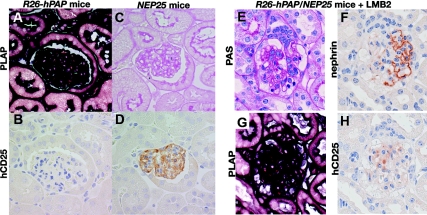
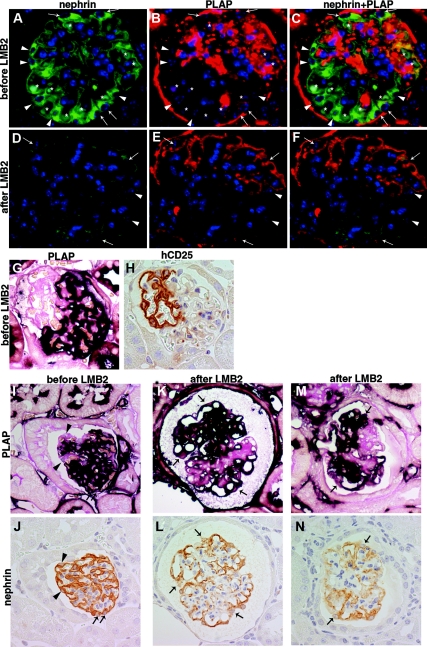
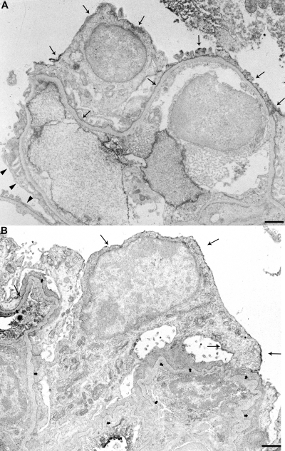
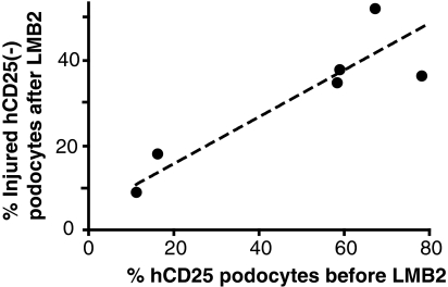
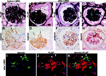
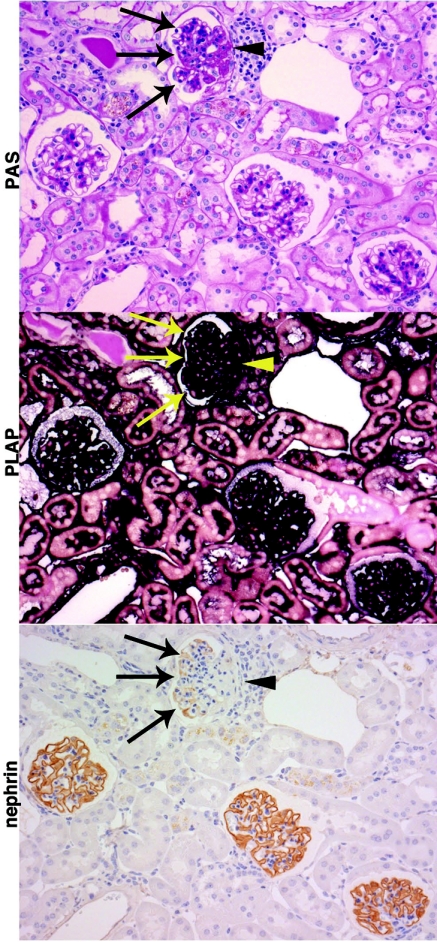
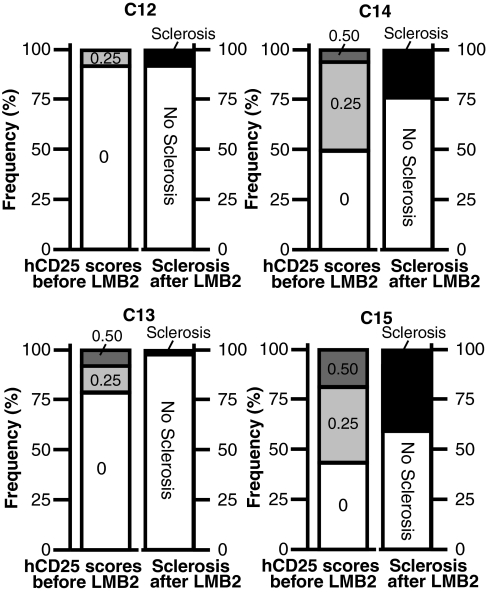
Loss of podocytes promotes glomerulosclerosis, but whether this results from a continued primary insult or a secondary mechanism triggered by the initial loss of podocytes is unknown. We generated chimeric mice in which only a subpopulation of podocytes expressed hCD25, which is the receptor for the immunotoxin LMB2. In addition, genetic labeling of hCD25-negative cells with human placental alkaline phosphatase allowed the study of these two distinct podocyte populations. Administration of LMB2 did not cause podocyte injury in hCD25-negative control mice. In contrast, LMB2 severely damaged or sloughed off the subpopulation of hCD25-positive podocytes within the chimeric glomeruli. Moreover, hCD25-negative podocytes, which were immune to the initial toxin injury, developed injury as early as 4 d after LMB2 injection, evidenced by foot process effacement, upregulation of desmin, and downregulation of nephrin, podocin, and podocalyxin. Furthermore, the magnitude of secondary injury correlated with the magnitude of primary injury, supporting the concept of an amplified cascade of podocyte injury. In conclusion, podocyte damage can propagate injury by triggering secondary damage of "remnant" intact podocytes, even when the primary insult is short-lived. This transmission of podocyte injury may form a vicious cycle leading to accelerated podocyte deterioration and glomerulosclerosis.


Figures

Figure 1.
Figure 1.
PLAP and hCD25 staining in R26-hPAP, NEP25, and R26-hPAP/NEP25 dual transgenic mice. (A through D): Kidneys of R26-hPAP and NEP25 mice were embedded in the same paraffin block and stained for PLAP and hCD25. In R26-hPAP mice, all types of cells are intensely stained for PLAP (A), but no cell for hCD25 (B). In NEP25 mice, no cell stain for PLAP (C), but all podocytes stain for hCD25. PLAP staining is most intense along the plasma membrane. (E through H) Podocyte injury was induced in R26-hPAP/NEP25 dual transgenic mice by injecting LMB2 (6 d after 25 ng/g BW of LMB2). The glomerulus shows vacuolar degeneration of epithelial cells, adhesion, and early sclerosis (E), segmental loss of nephrin staining (F), and global loss of hCD25 staining (H). In the same glomerulus, all cells are intensely stained for PLAP (G), thus demonstrating stable labeling of injured podocytes. A and C, B and D are from the same section. A and B, C and D, E through H are from adjacent sections. Magnification, ×400.
Figure 2.
Figure 2.
NEP25R26-hPAP chimeric mice at baseline and 4 d after 25 ng/g BW of LMB2. (A through F) Double immunofluorescent staining for nephrin (green) and PLAP (red) in a chimeric mouse before LMB2 (A through C) demonstrates global staining of nephrin and patchy (i.e., chimeric-pattern) staining of PLAP. Asterisks show RBCs with autofluorescence. Arrows depict DAPI nuclear staining (blue) with nephrin and PLAP, i.e., hCD25 (-) podocytes, and arrow heads depict DAPI with nephrin alone, i.e., hCD25 (+) podocytes. After LMB2 (D through F), nephrin staining is diminished globally, both in PLAP (+) podocytes (arrows) and in PLAP (-) podocytes (arrow heads), demonstrating nephrin is downregulated in hCD25 (-) podocytes. These panels and those in Figure S1 were stained in an identical fashion at the same time and photographed under the same conditions. (G and H) Serial sections from a NEP25R26-hPAP chimeric mouse at baseline were stained for PLAP and hCD25. PLAP (+) cells are negative for hCD25, and PLAP (-) surface cells are positive for hCD25. (I through N) Serial section analysis for PLAP and nephrin. I and J, K and L, M and N are from adjacent sections. Before LMB2 (I and J), both PLAP (+) (arrows) and PLAP(-)(arrow heads) surface podocytes show normal nephrin staining pattern. After LMB2 (K and L), nephrin staining is downregulated in some PLAP (+) surface podocytes (arrows) in both glomeruli with (M and N) and without (K and L) adhesion. Magnification, ×400.
Figure 3.
Figure 3.
Immunoelectronmicroscopy for PLAP in NEP25R26-hPAP chimeric glomerulus at baseline (A) and 9 d after injection of LMB2 (B). (A) Plasma membrane of the cell body and foot processes (arrows) and invaginated membrane of some podocytes are clearly stained with PLAP, whereas those of other podocytes (arrow heads) are not stained. (B) Plasma membranes (arrows) of most podocytes are intensely stained for PLAP (long arrows). The invaginated membrane is also stained for PLAP. The podocytes show extensive foot process effacement (short arrows) and microvillous transformation (asterisks). Bars represent 1 μm.
Figure 4.
Figure 4.
Correlation between hCD25 index before LMB2 and the average percentage of injured surface hCD25(-) podocytes after LMB2. hCD25 index, determined semiquantitatively, is the same as that shown in Table 1. The average percentage of injured surface hCD25(-) podocytes was determined as described in the Concise Methods section. Thus, the greater the number of hCD25(+) podocytes, which are the target of the primary injury, the greater the number of injured hCD25(-) podocytes that acquire secondary injury.
Figure 5.
Figure 5.
Injury in hCD25 (-) podocytes of NEP25R26-hPAP chimeric mice 9 d after 2.5 ng/g BW of LMB2. (A through H) Serial section analysis. Upper and lower panels are paired and from adjacent sections. Before LMB2 (A and B), both PLAP (+) (arrows) and PLAP(-)(arrow heads) surface podocytes show normal nephrin staining pattern. After LMB2, nephrin (A and D) and podocin (E and F) are downregulated and desmin (G and H) is upregulated in some PLAP (+) surface podocytes (arrows). (I through K) Double immunofluorescent staining for desmin and PLAP. Desmin is upregulated both in PLAP (+) podocytes (arrows) and in PLAP (-) podocytes (arrow heads), demonstrating damage of hCD25 (-) podocytes. Of note, PLAP (+) cells do not carry the hCD25 transgene and therefore are not targeted by LMB2. Magnification, ×400.
Figure 6.
Figure 6.
Focal segmental glomerulosclerosis in chimeric mice 6 wk after 25 ng/g BW of LMB2. Chimeric mice with relatively small baseline hCD25(+) podocyte population were injected with 25 ng/g BW of LMB2 and analyzed 6 wk later (mice C12, 13, 14, 15). Representative PAS, PLAP, and nephrin staining on serial sections from a representative mouse are shown. One glomerulus shows segmental sclerosis on PAS staining. All cells in the sclerotic lesion (arrow head) or in the nonsclerotic portion (arrows) are positive for PLAP, indicating that all hCD25(+) podocytes have been effectively eliminated by LMB2. The nonsclerotic portion shows extensive downregulation of nephrin. The other three glomeruli show normal structure in PAS staining and normal nephrin expression pattern. Magnification, ×200.
Figure 7.
Figure 7.
Frequency distribution of hCD25 scores before LMB2 (left columns) and the percentage of normal and sclerotic glomeruli after LMB2 (right columns). Each panel represents results from an individual mouse. The left columns represent frequency distribution of baseline hCD25 positivity and the right columns represent the percentage of normal and sclerotic glomeruli after LMB2. The former was determined on the basis of >55 glomeruli in a single section, and the latter was determined by serial section analysis on >70 glomeruli. hCD25 scores 0.00, 0.25, and 0.5 represent hCD25 areas of 0, 1 to 25, and 26 to 50%, respectively. None of these mice had even a single glomerulus with hCD25(+) area greater than 50% before LMB2, indicating that chimeric contribution of LMB2-susceptible podocytes was small in these mice. Mice C14 and C15 had greater number of glomeruli with LMB2-susceptible podocytes than mice C12 and C13, and later showed greater number (25.8 to 41.8%) of sclerotic glomeruli than C12 and C13 did (1 to 8.5%). To determine the threshold percentage of LMB2-susceptible podocytes that is sufficient to develop sclerosis, the two columns obtained from independent analyses are compared based on the notions (1) that the percentage of hCD25 (+) podocytes is similar between biopsied and autopsied kidneys of the same chimeric mouse, as demonstrated earlier, and (2) that glomeruli with more hCD25 (+) podocytes are more prone to develop glomerulosclerosis, which was shown at the whole kidney level. According to this limited analysis, threshold hCD25 positivity for sclerosis is variable among mice and is affected by percentage of glomeruli containing hCD25 (+) podocytes. Thus, mice C12 and C13 had the fewest glomeruli containing hCD25(+) podocytes and showed much less sclerosis than mice C14 and C15 with widespread hCD25(+) podocytes.

Comment in

  • Podocyte injury can be catching.
    D'Agati V. D'Agati V. J Am Soc Nephrol. 2011 Jul;22(7):1181-3. doi: 10.1681/ASN.2011050486. Epub 2011 Jun 16. J Am Soc Nephrol. 2011. PMID: 21680648 No abstract available.

Similar articles

Cited by

References

    1. LeHir M, Kriz W: New insights into structural patterns encountered in glomerulosclerosis. Curr Opin Nephrol Hypertens 16: 184–191, 2007 - PubMed
    1. D'Agati VD: Podocyte injury in focal segmental glomerulosclerosis: Lessons from animal models (a play in five acts). Kidney Int 73: 399–406, 2008 - PubMed
    1. Wiggins RC: The spectrum of podocytopathies: A unifying view of glomerular diseases. Kidney Int 71: 1205–1214, 2007 - PubMed
    1. Barisoni L, Schnaper HW, Kopp JB: A proposed taxonomy for the podocytopathies: A reassessment of the primary nephrotic diseases. Clin J Am Soc Nephrol 2: 529–542, 2007 - PubMed
    1. Mundel P, Shankland SJ: Podocyte biology and response to injury. J Am Soc Nephrol 13: 3005–3015, 2002 - PubMed

Publication types

Substances