

Mitral insufficiency due to an isolated anterior-leaflet cleft: correction with an autologous pericardial patch supported by artificial chordae
- PMID: 21720461
- PMCID: PMC3113113
Mitral insufficiency due to an isolated anterior-leaflet cleft: correction with an autologous pericardial patch supported by artificial chordae
Abstract
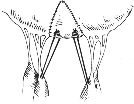
Congenital clefts of the mitral valve without an associated atrioventricular canal defect are rare, and they may cause mitral insufficiency that requires surgical correction. Repair is typically by direct suture; however, if the cleft is especially wide, the use of this technique may distort the valve leaflet and cause poor coaptation with valvular insufficiency.Herein, we present the case of a 39-year-old woman who had severe mitral valve insufficiency secondary to a wide isolated cleft of the anterior mitral leaflet. The valve was reconstructed with an autologous pericardial patch supported by polytetrafluoroethylene neochordae and an implanted annuloplasty ring. Echocardiographic examination 1 year postoperatively showed excellent competence of the mitral valve and good coaptation of the leaflets. To our knowledge, this is the 1st report that describes the use of artificial neochordae to support an autologous pericardial patch in the repair of a cleft in the anterior mitral valve leaflet.
Keywords: Adult; cardiac surgical procedures; echocardiography; heart defects, congenital/complications/surgery; mitral valve insufficiency/etiology/surgery; mitral valve/abnormalities/pathology/surgery; surgical techniques; treatment outcome.
Figures


Similar articles
-
Midterm Results of Mitral Valve Repair With Pericardial Leaflet Augmentation: A Single-Center Experience.Semin Thorac Cardiovasc Surg. 2020 Autumn;32(3):433-440. doi: 10.1053/j.semtcvs.2019.10.019. Epub 2019 Dec 19. Semin Thorac Cardiovasc Surg. 2020. PMID: 31863832
-
Cleft posterior mitral valve leaflet in an adult with Turner syndrome diagnosed with the use of 3-dimensional transesophageal echocardiography.Tex Heart Inst J. 2012;39(4):547-9. Tex Heart Inst J. 2012. PMID: 22949775 Free PMC article.
-
Leaflet-to-Annuloplasty Ring Clipping for Severe Mitral Regurgitation.JACC Cardiovasc Interv. 2016 Apr 11;9(7):e63-4. doi: 10.1016/j.jcin.2015.12.272. Epub 2016 Mar 4. JACC Cardiovasc Interv. 2016. PMID: 26952908 No abstract available.
-
The posterior mitral leaflet overhang: A rare yet possible complication of percutaneous mitral valve procedures.Cardiovasc Revasc Med. 2025 Feb;71:50-54. doi: 10.1016/j.carrev.2024.09.017. Epub 2024 Oct 15. Cardiovasc Revasc Med. 2025. PMID: 39426904 Review.
-
Optimizing P2 Neochordal Length and Stability in Mitral Valve Repair With Use of a Polypropylene Loop.Tex Heart Inst J. 2020 Jun 1;47(3):207-209. doi: 10.14503/THIJ-18-6913. Tex Heart Inst J. 2020. PMID: 32997772 Free PMC article. Review.
Cited by
-
Totally Endoscopic Repair of Isolated Congenital Anterior Mitral Leaflet Cleft: A Case Report.Innovations (Phila). 2025 Mar-Apr;20(2):217-218. doi: 10.1177/15569845251324488. Epub 2025 May 4. Innovations (Phila). 2025. PMID: 40320633 Free PMC article. No abstract available.
References
-
- Timoteo A, Galrinho A, Fiarresga A, Branco L, Banazol N, Leal A, et al. Isolated cleft of the anterior mitral valve leaflet. Eur J Echocardiogr 2007;8(1):59–62. - PubMed
-
- Perier P, Clausnizer B. Isolated cleft mitral valve: valve reconstruction techniques. Ann Thorac Surg 1995;59(1):56–9. - PubMed
-
- Edwards JE, Dry TJ, Parker RL, Burchell HB, Wood EH, Bulbulian AH. An atlas of congenital anomalies of the heart and great vessels. Springfield (IL): Charles C. Thomas; 1954. p. 41–2.
Publication types
MeSH terms
LinkOut - more resources
Full Text Sources
Medical