. 2011;6(6):e18919.
doi: 10.1371/journal.pone.0018919. Epub 2011 Jun 20.

Structural and histone binding ability characterizations of human PWWP domains

Affiliations

Structural and histone binding ability characterizations of human PWWP domains

Hong Wu et al. PLoS One. 2011.

Abstract

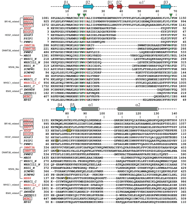
Background: The PWWP domain was first identified as a structural motif of 100-130 amino acids in the WHSC1 protein and predicted to be a protein-protein interaction domain. It belongs to the Tudor domain 'Royal Family', which consists of Tudor, chromodomain, MBT and PWWP domains. While Tudor, chromodomain and MBT domains have long been known to bind methylated histones, PWWP was shown to exhibit histone binding ability only until recently.

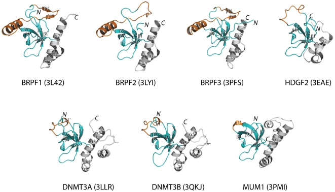
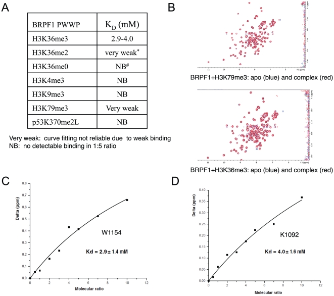
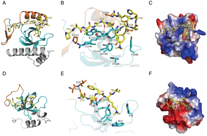
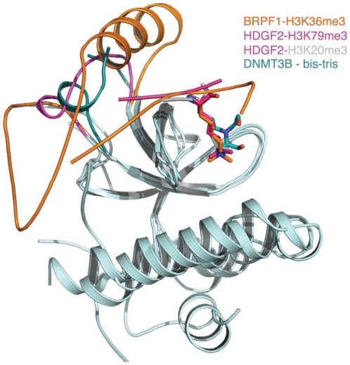
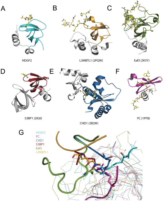
Methodology/principal findings: The PWWP domain has been shown to be a DNA binding domain, but sequence analysis and previous structural studies show that the PWWP domain exhibits significant similarity to other 'Royal Family' members, implying that the PWWP domain has the potential to bind histones. In order to further explore the function of the PWWP domain, we used the protein family approach to determine the crystal structures of the PWWP domains from seven different human proteins. Our fluorescence polarization binding studies show that PWWP domains have weak histone binding ability, which is also confirmed by our NMR titration experiments. Furthermore, we determined the crystal structures of the BRPF1 PWWP domain in complex with H3K36me3, and HDGF2 PWWP domain in complex with H3K79me3 and H4K20me3.

Conclusions: PWWP proteins constitute a new family of methyl lysine histone binders. The PWWP domain consists of three motifs: a canonical β-barrel core, an insertion motif between the second and third β-strands and a C-terminal α-helix bundle. Both the canonical β-barrel core and the insertion motif are directly involved in histone binding. The PWWP domain has been previously shown to be a DNA binding domain. Therefore, the PWWP domain exhibits dual functions: binding both DNA and methyllysine histones.

Enhanced version: This article can also be viewed as an enhanced version in which the text of the article is integrated with interactive 3D representations and animated transitions. Please note that a web plugin is required to access this enhanced functionality. Instructions for the installation and use of the web plugin are available in Text S1.


Conflict of interest statement

Competing Interests: The authors have declared that no competing interests exist.

Figures

Figure 1
Figure 1. Structure-based sequence alignment of human PWWP domains.
There are over 20 PWWP containing proteins in the human genome, which can be categorized into 6 classes. We solved crystal structures of PWWP domains from 7 different human proteins (colored in red and boxed). The structures solved by other labs are colored in red. All of these PWWP domains contain an aromatic cage formed by three aromatic residues (labeled by green triangles) except MBD5 and the N-terminal PWWP domain in NSD1. The identical residues in the alignment are colored in red. The secondary structure elements of BRPF1 are shown on top of the alignment, with cylinders representing helixes and arrows representing strands. The alignment was generated by using Clustal W assisted with manual adjustment.
Figure 2
Figure 2. Crystal structures of the seven human PWWP domains reported in this study.
PWWP fold consists of three structural elements: the canonical core (colored in cyan) comprising 5 β-strands, which highly resembles the Tudor domain fold, an insertion motif between the second and third β strands (colored in orange) and the PWWP characteristic C-terminal α helix motif consisting of 1–5 α-helixes (colored in grey). The figure was generated by Pymol.
Figure 3
Figure 3. NMR titration confirms that BRPF1 preferentially binds tri-methylated H3K36.
(A) Binding affinities of the BRPF1 PWWP domain to different histone peptides. (B)Superposition of 15N-1H HSQC NMR spectra of the BRPF1 PWWP domain in the presence (red) and absence (blue) of H3K79me3 (1∶10) and H3K36me3 (1∶10) peptides. (C) Binding affinity calculation based on the change in the chemical shifts of W1154 in the 15N-1H BRPF1 resonances upon addition of nonlabeled H3K36me3 peptide. (D) Binding affinity calculation based on the change in the chemical shifts of K1092 in the 15N-1H BRPF1 resonances upon addition of nonlabeled H3K36me3 peptide. The concentration of 15N BRPF1 in all NMR experiments is 0.2 mM. W1154 and K1092 are two residues from the BRPF1 PWWP domain.
Figure 4
Figure 4. Complex structures of BRPF1-H3K36me3 and HDGF2-H3K79me3.
(A) Structure of BRPF1 PWWP domain in complex H3K36me3 peptide. The PWWP domain is shown in cartoon, and the peptide is shown in a stick model. (B) The detailed interactions between the BRPF1 PWWP domain and H3K36me3. Hydrogen bonds are shown in dashed lines. (C) Electrostatic surface representation of BRPF1-H3K36me3 complex. (D) Structure of HDGF2 PWWP domain in complex H3K79me3 peptide. The PWWP domain is shown in cartoon, and the peptide is shown in a stick model. (E) The detailed interactions between the HDGF2 PWWP domain and H3K79me3. Hydrogen bonds are shown in dashed lines. (F) Electrostatic surface representation of HDGF2-H3K79me3 complex.
Figure 5
Figure 5. PWWP domains of DNMT3A and DNMT3B bind a bis-tris molecule in their respective aromatic cage.
(A) The detailed interactions between DNMT3A and a bis-tris molecule. (B) The detailed interactions between DNMT3B and a bis-tris molecule. The bis-tris molecule is shown in a green stick model, and the interacting residues from the PWWP domain are also shown in stick models. Hydrogen bonds are shown in dashed lines.
Figure 6
Figure 6. The peptides share a similar binding mode in the complex structures of BRPF1-H3K36me3, HDGF2-H3K79me3 and HDGF2-H4K20me3.
The bis-tris molecules in the DNMT3A/DNMT3B complex structures are bound at the same site as the tri-methyl-ammonium group of the methyllysine (DNMT3A structure is not shown here for clarity). The insertion motifs from these three PWWP domains may play a role in conferring the ligand specificity.
Figure 7
Figure 7. Comparison of the binding modes of methylated histone peptides to PWWP domain, chromo domain, Tudor domain and MBT repeat domain.
(A–F) Different histone code reader domains shown individually in complex with their corresponding ligands: HDGF2-H3K36me3 (cyan, A), L3MBTL1-H4K20me2 (yellow, B), Eaf3-a mimic H3K36me2 peptide (green, C), 53BP1-H4K20me2 (red, D), CHD1-H3K4me3 (blue, E), Polycomb-H3K27me3 (magenta, F). The peptides are shown in stick models. (G) Superposition of different histone code “reader” domains in complex with their corresponding ligands. All the structures are shown in the same orientation as they are in Fig. 7A–7F. The conserved β-sheet core among these domains is colored the same as in Fig. 7A–7F.

References

    1. Stec I, Nagl SB, van Ommen GJ, den Dunnen JT. The PWWP domain: a potential protein-protein interaction domain in nuclear proteins influencing differentiation? FEBS Lett. 2000;473:1–5. - PubMed
    1. Shikauchi Y, Saiura A, Kubo T, Niwa Y, Yamamoto J, et al. SALL3 interacts with DNMT3A and shows the ability to inhibit CpG island methylation in hepatocellular carcinoma. Mol Cell Biol. 2009;29:1944–1958. - PMC - PubMed
    1. Park J, Kim TY, Jung Y, Song SH, Kim SH, et al. DNA methyltransferase 3B mutant in ICF syndrome interacts non-covalently with SUMO-1. J Mol Med. 2008;86:1269–1277. - PubMed
    1. Qiu C, Sawada K, Zhang X, Cheng X. The PWWP domain of mammalian DNA methyltransferase Dnmt3b defines a new family of DNA-binding folds. Nat Struct Biol. 2002;9:217–224. - PMC - PubMed
    1. Chen T, Tsujimoto N, Li E. The PWWP domain of Dnmt3a and Dnmt3b is required for directing DNA methylation to the major satellite repeats at pericentric heterochromatin. Mol Cell Biol. 2004;24:9048–9058. - PMC - PubMed

Publication types

LinkOut - more resources