. 2011;6(6):e21587.
doi: 10.1371/journal.pone.0021587. Epub 2011 Jun 24.

Phenotypic expression of ADAMTS13 in glomerular endothelial cells

Affiliations

Phenotypic expression of ADAMTS13 in glomerular endothelial cells

Ramesh Tati et al. PLoS One. 2011.

Abstract

Background: ADAMTS13 is the physiological von Willebrand factor (VWF)-cleaving protease. The aim of this study was to examine ADAMTS13 expression in kidneys from ADAMTS13 wild-type (Adamts13⁺/⁺) and deficient (Adamts13⁻/⁻) mice and to investigate the expression pattern and bioactivity in human glomerular endothelial cells.

Methodology/principal findings: Immunohistochemistry was performed on kidney sections from ADAMTS13 wild-type and ADAMTS13-deficient mice. Phenotypic differences were examined by ultramorphology. ADAMTS13 expression in human glomerular endothelial cells and dermal microvascular endothelial cells was investigated by real-time PCR, flow cytometry, immunofluorescence and immunoblotting. VWF cleavage was demonstrated by multimer structure analysis and immunoblotting. ADAMTS13 was demonstrated in glomerular endothelial cells in Adamts13⁺/⁺ mice but no staining was visible in tissue from Adamts13⁻/⁻ mice. Thickening of glomerular capillaries with platelet deposition on the vessel wall was detected in Adamts13⁻/⁻ mice. ADAMTS13 mRNA and protein were detected in both human endothelial cells and the protease was secreted. ADAMTS13 activity was demonstrated in glomerular endothelial cells as cleavage of VWF.

Conclusions/significance: Glomerular endothelial cells express and secrete ADAMTS13. The proteolytic activity could have a protective effect preventing deposition of platelets along capillary lumina under the conditions of high shear stress present in glomerular capillaries.


Conflict of interest statement

Competing Interests: The authors have declared that no competing interests exist.

Figures

Figure 1
Figure 1. ADAMTS13 expression in mouse kidney.
ADAMTS13 expression was investigated by immunohistochemistry in mouse renal tissue. Staining was observed in the kidney of Adamts13+/+ wild-type mice on the mixed 129X1/SvJ and C57BL/6 genetic background (Panel A). The inset in panel A shows glomerular capillary staining (arrow) at a higher magnification. Tissue from Adamts13−/− mice (same genetic background) did not label for ADAMTS13 (Panel B). Reproducible results were obtained in six separate experiments including mice with the mixed C57BL/6J and CAST/Ei genetic background. Images are at 1000x magnification. P: podocyte, US: urinary space, T: tubular cell.
Figure 2
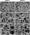
Figure 2. Ultramorphology of glomeruli in wild-type and deficient mice.
Scanning electron microscopy of kidney from wild-type mice showed normal morphology as depicted in panel A. In the renal glomeruli the Bowman's capsule (BC), glomerular basement membrane (GBM), endothelial cell capillaries (C), erythrocytes (E), podocytes (P) and urinary space (US) were identified. Panel B shows a glomerulus from an Adamts13−/− mouse demonstrating thickened and irregular capillary walls (arrow). These findings were enlarged in panel C from another glomerulus from an Adamts13+/+ mouse showing thin glomerular capillary walls (arrow heads) and in panel D from an Adamts13−/− mouse showing thickened capillary walls (arrows) with deposits on the vessel lumina. Panel E demonstrates lack of platelet deposits in glomerular capillaries from an Adamts13+/+mouse, further confirmed by immunoelectron microscopy (insets). Panel F was taken from another glomerulus in an Adamts13−/− mouse and shows platelets (within circles) deposited on a vessel wall. Insets in panel F show immunoelectron microscopy with labeled platelets (arrows) deposited on glomerular capillary walls. Scale bar represents 10 µm (A, B), 5 µm (C–F) and 2.5 µm (insets in E, F). Panels A, B, D–F and insets were from mice with the 129X1/SvJ and C57BL/6 genetic background and panel C was from mice with the C57BL/6J and CAST/Ei genetic background.
Figure 3
Figure 3. ADAMTS13 mRNA expression in cultured endothelial cells.
ADAMTS13 gene transcripts in endothelial cells were detected by real-time PCR. A. Using the probe against exons 28–29 the amplification plot indicates detectable ADAMTS13 mRNA levels in CiGEnC and HMVEC cells. Human kidney (HK) and human liver (HL) were used as the positive controls. No template control (NTC) and CHO cells were used as the negative controls and showed no amplification. B. The housekeeping gene 18S, which was expressed at comparable levels in all samples, was used to standardize the data. The upper limit of the boxes depicts the means and bars are standard error of the mean from three separate cell culture experiments.
Figure 4
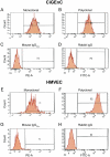
Figure 4. Detection ofADAMTS13 in cultured endothelial cells by flow cytometry.
ADAMTS13 protein was detected in CiGEnC (Panels A–D) using A10 monoclonal antibody (panel A) and SU19 polyclonal antibody (panel B). The control antibodies, mouse IgG2b-κ (panel C) and rabbit IgG (panel D) did not bind to any cells. HMVEC showed similar results (Panels E–H). Reproducible results were obtained from two different experiments.
Figure 5
Figure 5. ADAMTS13 expression detected intracellularly by immunofluorescence.
ADAMTS13 protein expression was investigated in CiGEnC (panels A–B) and HMVEC (panels C–D). ADAMTS13 was detected in CiGEnC using SU19 polyclonal antibody (panel A). The SU19 antibody was pre-incubated with blocking peptide resulting in marked signal reduction (panel B) in which FITC labeling of ADAMTS13 was abolished and blue DAPI labeling marked cell nuclei. HMVEC cells showed similar results staining positively for ADAMTS13 with the polyclonal SU19 antibody (C). SU19 blocking experiments resulted in marked decrease in signal intensity (D). Reproducible results were achieved in at least six separate cell experiments from three different passages. All images are at 400x magnification.
Figure 6
Figure 6. ADAMTS13 is secreted into the media.
Recombinant ADAMTS13 showed a band at 150 kDa under non-reducing conditions (lane 1, the positive control) and control medium did not show any band (lane 2). Media from CiGEnC and HMVEC exhibited similar bands at approximately 150 kDa (lanes 3 and 4, respectively). Reproducible results were obtained from four different experiments.
Figure 7
Figure 7. ADAMTS13 activity depicted by the VWF multimer structure.
Endothelial cell lysates were incubated with purified VWF. Cell buffer incubated with VWF was used as the negative control and showed high molecular weight VWF multimers (lanes 1 and 3). CiGEnC and HMVEC lysates showed marked cleavage of VWF multimers (lanes 2 and 4, respectively). The two panels were run on separate gels but within each panel samples were run on the same gel. Reproducible results were obtained from three separate experiments.
Figure 8
Figure 8. VWF cleavage by endothelial cell lysates depicted by immunoblotting.
A. Recombinant ADAMTS13 cleaves the A2 domain of VWF whereby 176 kDa and 140 kDa bands appear (lane 1). Full-length VWF is depicted by an arrow. Cell buffer, incubated with VWF and used as the negative control, did not exhibit the cleavage fragments (lane 2). CiGEnC cell lysate exhibited VWF cleaving activity as shown by the presence of similar cleavage fragments (lane 3) which were inhibited by 20 mM EDTA (lane 4). HMVEC lysate also showed the two breakdown products (lane 5) which were partially inhibited by EDTA (lane 6). B. Immunoadsorption of ADAMTS13 inhibited VWF cleaving activity. Recombinant ADAMTS13 showed the two cleavage fragments (lane 1) and the cleaving activity was abrogated by removal of ADAMTS13 (lane 2). CiGEnC cell lysate also induced VWF cleavage (lane 3) which was inhibited by immunoadsorption of ADAMTS13 from the lysate (lane 4). Experiments were carried out three times with reproducible results. C. VWF cleavage products were derived from exogenous VWF. Exogenous VWF was added to rADAMTS13 resulting in two cleavage products (lane 1). CiGEnC lysate, without added exogenous VWF, did not show cleavage bands but exhibited a weak band corresponding to full-length VWF (lane 2). When purified exogenous VWF was added to CiGEnC lysates VWF cleavage was demonstrated (lane 3). D. Recombinant ADAMTS13 cleaves exogenously added VWF in cell medium (that was not in contact with endothelial cells, lane 1). Cell medium (that had not been exposed to cells) without added ADAMTS13 exhibited full-length VWF and a very weak band at 176 kDa (lane 2). Cell medium derived from unstimulated CiGEnC exhibited a stronger band at 176 kDa and a weak band at 140 kDa (lane 3). Cell medium derived from CiGEnC stimulated with estradiol exhibited a band at 176 kDa and a weak band at 140 kDa (lane 4). All lanes were run on the same gel. Experiments were carried out twice with reproducible results.
Figure 9
Figure 9. Lack of detectable MMP9 in the endothelial cells.
MMP9 protein was not detected in either CiGEnC (panel A) or HMVEC (panel B) using mouse anti-human MMP9 antibody investigated by flow cytometry. The control antibody was negative (panels C and D). Results were reproduced in two separate experiments.

References

    1. Furlan M, Robles R, Lamie B. Partial purification and characterization of a protease from human plasma cleaving von Willebrand factor to fragments produced by in vivo proteolysis. Blood. 1996;87:4223–4234. - PubMed
    1. Moake JL. von Willebrand factor in thrombotic thrombocytopenic purpura. Blood. 1986;67:1523–1526. - PubMed
    1. Jaffe EA, Hoyer LW, Nachman RL. Synthesis of von Willebrand factor by cultured human endothelial cells. Proc Natl Acad Sci U S A. 1974;71:1906–1909. - PMC - PubMed
    1. Sporn LA, Chavin SI, Marder VJ, Wagner DD. Biosynthesis of von Willebrand protein by human megakaryocytes. J Clin Invest. 1985;76:1102–1106. - PMC - PubMed
    1. Tsai HM, Nagel RL, Hatcher VB, Sussman, II Multimeric composition of endothelial cell-derived von Willebrand factor. Blood. 1989;73:2074–2076. - PubMed

Publication types

MeSH terms