

Stress response gene activation protects sea urchin embryos exposed to X-rays
- PMID: 21720812
- PMCID: PMC3220391
- DOI: 10.1007/s12192-011-0277-3
Stress response gene activation protects sea urchin embryos exposed to X-rays
Abstract
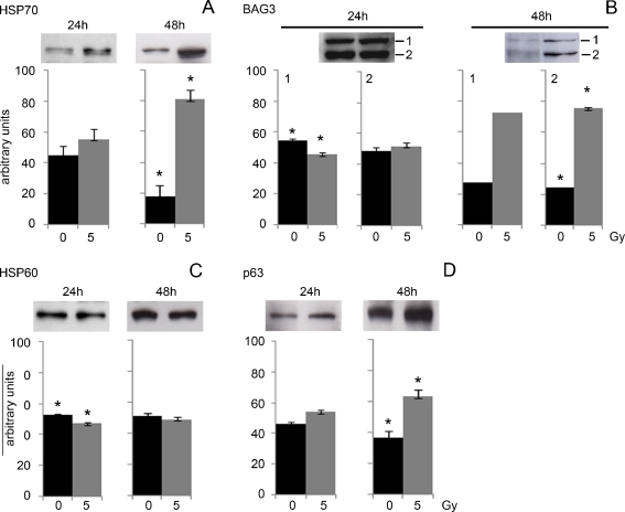
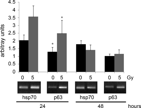
We used Paracentrotus lividus sea urchin embryos, a well-established model in developmental biology and ecotoxicology, for investigation on stress/anti-apoptotic protein expression elicited in response to harmful ionizing radiation, such as X-rays. We evaluated the acute effects of a high-dose exposure (5 Gy) on P. lividus analyzing by Western blotting the accumulation levels of HSP60, HSP70, BAG3 and a putative p63 at 24 and 48 h after irradiation. We found an increase in the HSP70, BAG3, and p63 protein levels only 48 h after irradiation, whereas no HSP60 increase was detected either at 24 or 48 h. Levels of the mRNA coding for HSP70 and p63 were also investigated by relative RT-PCR and were found to increase 24 h after irradiation, returning to their initial levels at 48 h. Results demonstrate the presence of an adaptive regulatory mechanism operating at the transcriptional level at 24 h, followed by a translational activation at 48 h post-irradiation. In conclusion, our findings confirm the sea urchin embryo as a sensible bioindicator of cell damage and we propose this model for studies on the protective pathways activated in response to X-rays. The novel result of the involvement of BAG3 and p63 in the response to X-rays, never tested so far in any other embryonic system, opens the way for their use as biomarkers of X-ray hazards.
Figures
Similar articles
-
Embryonic development and skeletogenic gene expression affected by X-rays in the Mediterranean sea urchin Paracentrotus lividus.Ecotoxicology. 2010 Mar;19(3):530-7. doi: 10.1007/s10646-009-0444-9. Epub 2009 Nov 27. Ecotoxicology. 2010. PMID: 19943107
-
Transcriptional increase and misexpression of 14-3-3 epsilon in sea urchin embryos exposed to UV-B.Cell Stress Chaperones. 2010 Nov;15(6):993-1001. doi: 10.1007/s12192-010-0210-1. Epub 2010 Jul 4. Cell Stress Chaperones. 2010. PMID: 20607471 Free PMC article.
-
Amino-modified polystyrene nanoparticles affect signalling pathways of the sea urchin (Paracentrotus lividus) embryos.Nanotoxicology. 2017 Mar;11(2):201-209. doi: 10.1080/17435390.2017.1279360. Epub 2017 Jan 24. Nanotoxicology. 2017. PMID: 28091127
-
Proteomic responses of sea urchin embryos to stressful ultraviolet radiation.Integr Comp Biol. 2012 Nov;52(5):665-80. doi: 10.1093/icb/ics058. Epub 2012 May 10. Integr Comp Biol. 2012. PMID: 22576820 Review.
-
Studies on heat shock proteins in sea urchin development.Dev Growth Differ. 1999 Aug;41(4):375-80. doi: 10.1046/j.1440-169x.1999.00450.x. Dev Growth Differ. 1999. PMID: 10466924 Review.
Cited by
-
Defensome against toxic diatom aldehydes in the sea urchin Paracentrotus lividus.PLoS One. 2012;7(2):e31750. doi: 10.1371/journal.pone.0031750. Epub 2012 Feb 20. PLoS One. 2012. PMID: 22363721 Free PMC article.
-
Molecular response to toxic diatom-derived aldehydes in the sea urchin Paracentrotus lividus.Mar Drugs. 2014 Apr 4;12(4):2089-113. doi: 10.3390/md12042089. Mar Drugs. 2014. PMID: 24714125 Free PMC article.
-
Gene Expression Analysis of the Stress Response to Lithium, Nickel, and Zinc in Paracentrotus lividus Embryos.Toxics. 2022 Jun 14;10(6):325. doi: 10.3390/toxics10060325. Toxics. 2022. PMID: 35736933 Free PMC article.
-
Maternal Exposure to Cadmium and Manganese Impairs Reproduction and Progeny Fitness in the Sea Urchin Paracentrotus lividus.PLoS One. 2015 Jun 30;10(6):e0131815. doi: 10.1371/journal.pone.0131815. eCollection 2015. PLoS One. 2015. PMID: 26125595 Free PMC article.
-
Elevated temperature attenuates ovarian functions and induces apoptosis and oxidative stress in the American oyster, Crassostrea virginica: potential mechanisms and signaling pathways.Cell Stress Chaperones. 2019 Sep;24(5):957-967. doi: 10.1007/s12192-019-01023-w. Epub 2019 Jul 30. Cell Stress Chaperones. 2019. PMID: 31363994 Free PMC article.
References
-
- Aluigi MG, Angelini C, Corte G, Falugi C. The sea urchin, Paracentrotus lividus, embryo as a “bioethical” model for neurodevelopmental toxicity testing: effects of diazinon on the intracellular distribution of OTX2-like proteins. Cell Biol Toxicol. 2008;24:587–601. doi: 10.1007/s10565-008-9061-2. - DOI - PubMed
Publication types
MeSH terms
Substances
LinkOut - more resources
Full Text Sources
Research Materials
Miscellaneous
