

doi: 10.1002/adma.201101516.
Epub 2011 Jul 1.
Nanofibrous lipid membranes capable of functionally immobilizing antibodies and capturing specific cells
Affiliations
- PMID: 21721057
- PMCID: PMC3175633
- DOI: 10.1002/adma.201101516
Item in Clipboard
Nanofibrous lipid membranes capable of functionally immobilizing antibodies and capturing specific cells
Adv Mater.
.
No abstract available
Figures
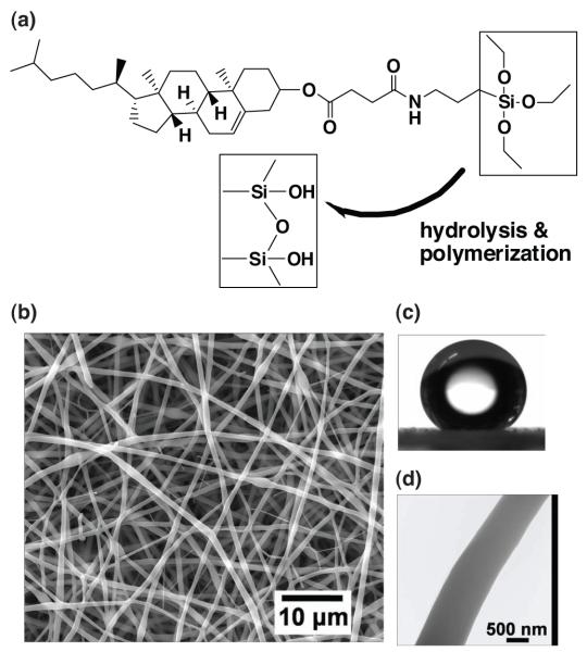
A combined sol-gel and electrospinning process for engineering nanofibrous CSS membrane. (a) The chemical structure of CSS and schematic of CSS hydrolysis and polymerization prior to electrospinning. (b) SEM images of electrospun CSS nanofibers. (c) Water contact angle images. (d) TEM images.
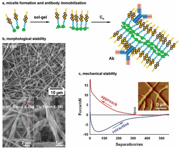
(a) Schematic of CSS polymerization and micelle formation. (b) SEM images of CSS fi bers that were immersed in 1× PBS for 72 h, 0.1% Triton X-100, and 1% Triton X-100 for 48 hrs. (c) A representative force-separation curve of single CSS fibers obtained via AFM nanoindentation. Inset: a typical AFM image of CSS fibers in the reflection mode.
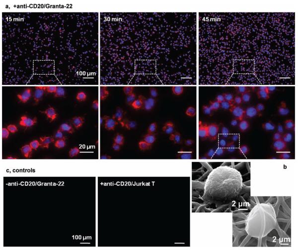
Immunofluorescent (a) and SEM (b) images of Granta-22 cells captured on the anti-CD20 mAb functionalized CSS membranes. The cells were fixed at indicated times, and stained for nucleus with DAPI (in blue) and for actin with phalloidin (in red). Inserts show the F-actin accumulated in the lamellipodias. Control studies (c): capturing no Granta-22 cells using the non-functionalized membranes, and no Jurkat T cells using the functionalized membranes in 45 min.
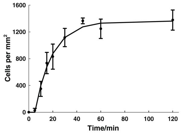
The number of the captured Granta-22 cells as a function of time. The solid line represents predictions from the first-order kinetic model, while symbols with error bars represent experimental data.
Similar articles
-
Combination anti-CD74 (milatuzumab) and anti-CD20 (rituximab) monoclonal antibody therapy has in vitro and in vivo activity in mantle cell lymphoma.Blood. 2011 Apr 28;117(17):4530-41. doi: 10.1182/blood-2010-08-303354. Epub 2011 Jan 12. Blood. 2011. PMID: 21228331 Free PMC article.
-
Immobilization of antibodies and enzymes on 3-aminopropyltriethoxysilane-functionalized bioanalytical platforms for biosensors and diagnostics.Chem Rev. 2014 Nov 12;114(21):11083-130. doi: 10.1021/cr5000943. Epub 2014 Oct 9. Chem Rev. 2014. PMID: 25299435 Review. No abstract available.
-
Efficient protein immobilization on polyethersolfone electrospun nanofibrous membrane via covalent binding for biosensing applications.Mater Sci Eng C Mater Biol Appl. 2016 Jan 1;58:586-94. doi: 10.1016/j.msec.2015.09.007. Epub 2015 Sep 6. Mater Sci Eng C Mater Biol Appl. 2016. PMID: 26478348
-
Micropatterns of double-layered nanofiber scaffolds with dual functions of cell patterning and metabolite detection.Lab Chip. 2011 Sep 7;11(17):2849-57. doi: 10.1039/c1lc20186g. Epub 2011 Jul 8. Lab Chip. 2011. PMID: 21738946
-
CD20-targeting in B-cell malignancies: novel prospects for antibodies and combination therapies.Invest New Drugs. 2016 Aug;34(4):497-512. doi: 10.1007/s10637-016-0349-4. Epub 2016 Apr 13. Invest New Drugs. 2016. PMID: 27075017 Review.
Cited by
-
Atomic force microscopy of electrospun organic-inorganic lipid nanofibers.Appl Phys Lett. 2011 Sep 5;99(10):103702-1037023. doi: 10.1063/1.3635783. Epub 2011 Sep 8. Appl Phys Lett. 2011. PMID: 21990942 Free PMC article.
-
A biomimetic mechanism for antibody immobilization on lipid nanofibers for cell capture.Appl Phys Lett. 2012 Nov 5;101(19):193701. doi: 10.1063/1.4766191. Epub 2012 Nov 6. Appl Phys Lett. 2012. PMID: 23213266 Free PMC article.
-
Comparative study of antibody immobilization mediated by lipid and polymer fibers.Colloids Surf B Biointerfaces. 2015 Oct 1;134:1-7. doi: 10.1016/j.colsurfb.2015.06.021. Epub 2015 Jun 19. Colloids Surf B Biointerfaces. 2015. PMID: 26141437 Free PMC article.
-
Centering of organic-inorganic hybrid liposomal cerasomes in electrospun gelatin nanofibers.Appl Phys Lett. 2012 Jan 16;100(3):33702-337023. doi: 10.1063/1.3678040. Epub 2012 Jan 18. Appl Phys Lett. 2012. PMID: 22312181 Free PMC article.
-
Lipid-mediated protein functionalization of electrospun polycaprolactone fibers.Express Polym Lett. 2016 May;10(5):430-437. doi: 10.3144/expresspolymlett.2016.40. Express Polym Lett. 2016. PMID: 32206095 Free PMC article.
References
-
- Simons K, Vaz WLC. Annu. Rev. Biophys. Biomol. Struct. 2004;33:269. - PubMed
- Han XJ, Studer A, Sehr H, Geissbuhler I, Di Berardino M, Winkler FK, Tiefenauer LX. Adv. Mater. 2007;19:4466.
-
- Lian T, Ho RJY. J. Pharm. Sci. 2001;90:667. - PubMed
-
- Shrout AL, Montefusco DJ, Weis RM. Biochemistry. 2003;42:13379. - PubMed
-
- McKee MG, Layman JM, Cashion MP, Long TE. Science. 2006;311:353. - PubMed
Publication types
MeSH terms
Substances
Grants and funding
LinkOut - more resources
Full Text Sources
Other Literature Sources
