. 2011 Jun;16(6):066014.
doi: 10.1117/1.3592499.

Spectrally resolving and scattering-compensated x-ray luminescence/fluorescence computed tomography

Affiliations

Spectrally resolving and scattering-compensated x-ray luminescence/fluorescence computed tomography

Wenxiang Cong et al. J Biomed Opt. 2011 Jun.

Abstract

The nanophosphors, or other similar materials, emit near-infrared (NIR) light upon x-ray excitation. They were designed as optical probes for in vivo visualization and analysis of molecular and cellular targets, pathways, and responses. Based on the previous work on x-ray fluorescence computed tomography (XFCT) and x-ray luminescence computed tomography (XLCT), here we propose a spectrally-resolving and scattering-compensated x-ray luminescence/fluorescence computed tomography (SXLCT or SXFCT) approach to quantify a spatial distribution of nanophosphors (other similar materials or chemical elements) within a biological object. In this paper, the x-ray scattering is taken into account in the reconstruction algorithm. The NIR scattering is described in the diffusion approximation model. Then, x-ray excitations are applied with different spectra, and NIR signals are measured in a spectrally resolving fashion. Finally, a linear relationship is established between the nanophosphor distribution and measured NIR data using the finite element method and inverted using the compressive sensing technique. The numerical simulation results demonstrate the feasibility and merits of the proposed approach.


Figures

Figure 1
Figure 1
Significance of x-ray scattering. (a) X-ray pencil beam passes through a water phantom and (b) 50% of scattered photons are distributed in the region delimited by the blue and green curves.
Figure 2
Figure 2
SXLCT system setup that uses x-rays under spectral modulation.
Figure 3
Figure 3
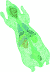
Mouse phantom represented in a finite element mesh.
Figure 4
Figure 4
Spatial resolution comparison between the reconstructed and true nanophosphor density distributions. (a) Reconstructed nanophosphor density distribution with the proposed SXLCT approach, yielding an average relative error of 10%; (b) reconstructed nanophosphor density distributions assuming no x-ray scattering in the model, yielding an average relative error over 40%; and (c) true nanophosphor density distribution.
Figure 5
Figure 5
Density resolution comparison between the reconstructed and true nanophosphor density distributions. (a) Reconstructed nanophosphor density distribution with the proposed SXLCT approach, producing an average relative error of 12%; (b) reconstructed nanophosphor density distribution assuming no x-ray scattering, resulting in an average relative error over 40%; and (c) true nanophosphor density distribution.
Figure 6
Figure 6
Spectral resolution comparison between the reconstructed and true nanophosphor density distributions. (a) Reconstructed distribution for nanophosphors of type A; (b) true distribution for nanophosphors of type A; (c) reconstructed distribution for nanophosphors of type B; and (d) true distribution for nanophosphors of type B.

References

    1. Minchin R. F. and Martin D. J., “Minireview: nanoparticles for molecular imaging—an overview,” Endocrinology 151, 474–481 (2010).10.1210/en.2009-1012 - DOI - PubMed
    1. Qian X., Peng X. H., Ansari D. O., Yin-Goen Q., Chen G. Z., Shin D. M., Yang L., Young A. N., Wang M. D., and Nie S., “In vivo tumor targeting and spectroscopic detection with surface-enhanced Raman nanoparticle tags,” Nat. Biotechnol. 26, 83–90 (2008).10.1038/nbt1377 - DOI - PubMed
    1. Tian Y., Cao W.-H., Luo X.-X., and Fu Y., “Preparation and luminescence property of Gd2O2S:Tb X-ray nano-phosphors using the complex precipitation method,” J. Alloys. Compd. 433, 313–317 (2007).10.1016/j.jallcom.2006.06.075 - DOI
    1. Xing M., Cao W., Pang T., Ling X., and Chen N., “Preparation and characterization of monodisperse spherical particles of X-ray nanophosphors based on Gd2O2S:Tb,” Chin. Sci. Bull. 54, 2982–2986 (2009).10.1007/s11434-009-0492-9 - DOI
    1. Carpenter C., Pratx G., Sun C., and Xing L., “Hybrid x-ray/optical luminescence imaging: characterization of experimental conditions,” Med. Phys. 37, 4011–4018 (2010).10.1118/1.3457332 - DOI - PMC - PubMed

Publication types

Substances