. 2011 Jun;6(2):43-53.
doi: 10.1116/1.3583535.

Modulation of fibroblast inflammatory response by surface modification of a perfluorinated ionomer

Affiliations

Modulation of fibroblast inflammatory response by surface modification of a perfluorinated ionomer

Thelma I Valdes et al. Biointerphases. 2011 Jun.

Abstract

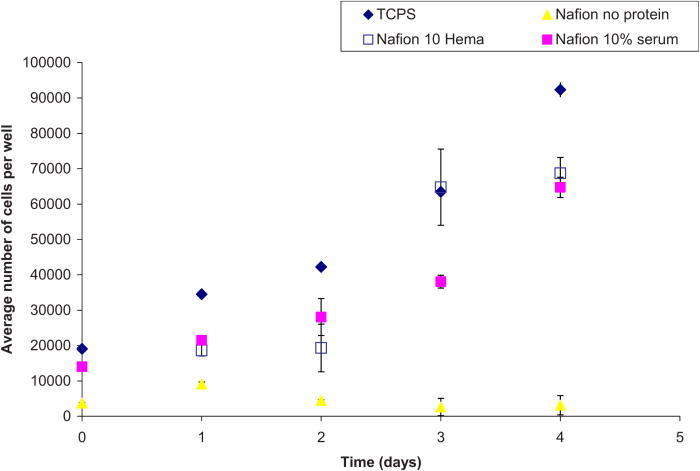
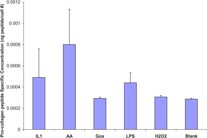
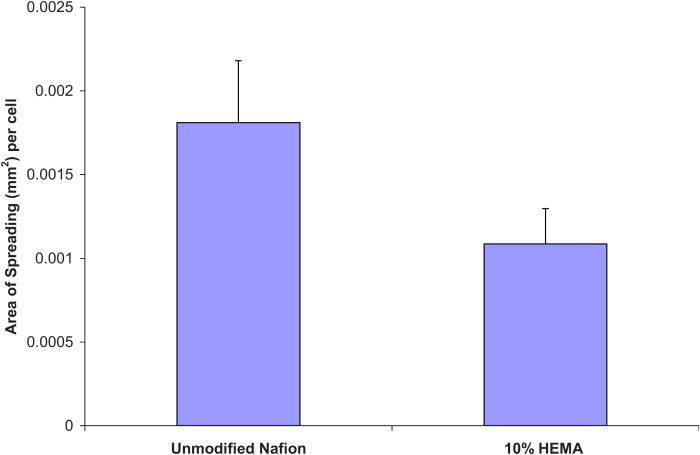
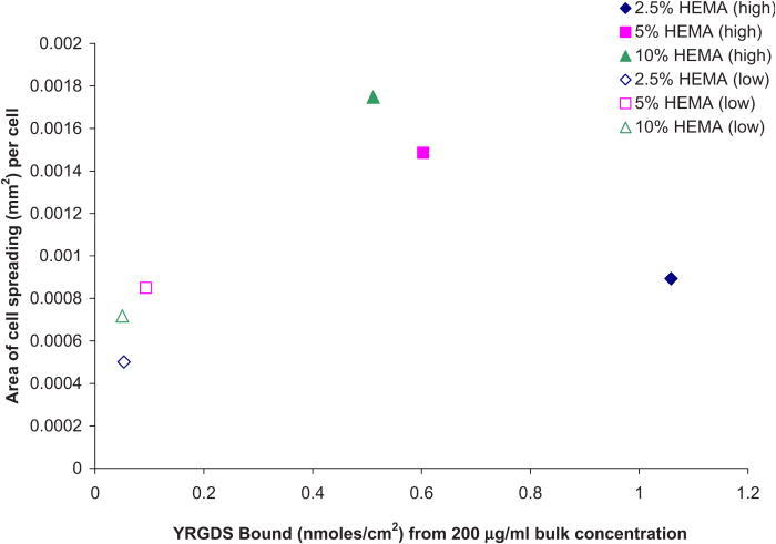
An ideal surface for implantable glucose sensors would be able to evade the events leading to chronic inflammation and fibrosis, thereby extending its utility in an in vivo environment. Nafion™, a perfluorinated ionomer, is the membrane material preferred for in situ glucose sensors. Unfortunately, the surface properties of Nafion™ promote random protein adsorption and eventual foreign body encapsulation, thus leading to loss of glucose signal over time. Details of the techniques to render Nafion™ nonprotein fouling are given in a previous article [T. I. Valdes et al., Biomaterials 29, 1356 (2008)]. Once random protein adsorption is prevented, a biologically active peptide can be covalently bonded to the treated Nafion™ to induce cellular adhesion. Cellular responses to these novel decorated Nafion™ surfaces are detailed here, including cell viability, cell spreading, and type I collagen synthesis. Normal human dermal fibroblasts (NHDFs) were cultured on control and modified Nafion™ surfaces. Findings indicate that Nafion™ modified with 10% 2-hydroxyethyl methacrylate and 90% tetraglyme created a nonfouling surface that was subsequently decorated with the YRGDS peptide. NHDFs were shown to have exhibited decreased type I collagen production in comparison to NHDF cells on unmodified Nafion™ surfaces. Here, the authors report evidence that proves that optimizing conditions to prevent protein adsorption and enhance cellular adhesion may eliminate fibrous encapsulation of an implant.


Figures

F<sc>ig</sc>. 1.
Fig. 1.
Adherent NHDF cells (per cm2) on unmodified Nafion surfaces preadsorbed with various proteins.
F<sc>ig</sc>. 2.
Fig. 2.
Typical cellular morphologies for NHDF on unmodified Nafion surfaces and TCPS.
F<sc>ig</sc>. 3.
Fig. 3.
Area of NHDF spreading quantified on unmodified Nafion surfaces preadsorbed with various proteins.
F<sc>ig</sc>. 4.
Fig. 4.
Adherent NHDF cells (per cm2) on various modified Nafion surfaces with two different bulk peptide concentrations (actual surface concentrations unknown; only bulk concentration reported; assumes a linear relationship).
F<sc>ig</sc>. 5.
Fig. 5.
Cells adhered on 10% HEMA surfaces with 200 μg / ml YRGDS vs number of cells plated.
F<sc>ig</sc>. 6.
Fig. 6.
Typical cellular morphologies for NHDF on modified Nafion surfaces at two different concentrations of YRGDS. Cells were allowed to adhere for 24 h.
F<sc>ig</sc>. 7.
Fig. 7.
Immunocytochemistry of NHDF on various substrata. “Merged” refers to an overlay of actin and paxillin photographs.
F<sc>ig</sc>. 8.
Fig. 8.
Area of cell spreading on various modified Nafion surfaces.
F<sc>ig</sc>. 9.
Fig. 9.
Proliferation assay for NHDF on various Nafion surfaces.
F<sc>ig</sc>. 10.
Fig. 10.
Procollagen ELISA on unmodified Nafion surfaces. Results reported as procollagen peptides per cell. Blank refers to serum-free media.
F<sc>ig</sc>. 11.
Fig. 11.
Procollagen ELISA on 10% HEMA modified Nafion surfaces. Results reported as procollagen peptides per cell. There was no detectable collagen in Gox or H2O2 conditions. Blank refers to serum-free media.
F<sc>ig.</sc> 12.
Fig. 12.
Area of cell spreading quantified after 4 days of being in culture in blank media.
F<sc>ig</sc>. 13.
Fig. 13.
Correlation between cell spreading and linked YRGDS at the various modified Nafion surfaces.

Similar articles

Cited by

References

    1. Daniloff G. Y., Diabetes Technol. Ther. 1, 261 (1999)10.1089/152091599317170. - DOI - PubMed
    1. Heller A., Annu. Rev. Biomed. Eng. 1, 153 (1999)10.1146/annurev.bioeng.1.1.153. - DOI - PubMed
    1. Chung T. D., Jeong R. A., Kang S. K. and Kim H. C., Biosens. Bioelectron. 16, 1079 (2001)10.1016/S0956-5663(01)00185-3. - DOI - PubMed
    1. Yang H., Chung T. D., Kim Y. T., Choi C. A., Jun C. H. and Kim H. C., Biosens. Bioelectron. 17, 251 (2002)10.1016/S0956-5663(01)00266-4. - DOI - PubMed
    1. Kang S. K., Jeong R. A., Park S., Chung T. D. and Kim H. C., Anal. Sci. 19, 1481 (2003)10.2116/analsci.19.1481. - DOI - PubMed

Publication types

MeSH terms