Review
. 2011:27:237-63.
doi: 10.1146/annurev-cellbio-092910-154251. Epub 2011 Jun 20.

Wound repair: toward understanding and integration of single-cell and multicellular wound responses

Affiliations
Review

Wound repair: toward understanding and integration of single-cell and multicellular wound responses

Kevin J Sonnemann et al. Annu Rev Cell Dev Biol. 2011.

Abstract

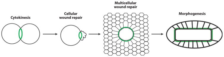
The importance of wound healing to medicine and biology has long been evident, and consequently, wound healing has been the subject of intense investigation for many years. However, several relatively recent developments have added new impetus to wound repair research: the increasing application of model systems; the growing recognition that single cells have a robust, complex, and medically relevant wound healing response; and the emerging recognition that different modes of wound repair bear an uncanny resemblance to other basic biological processes such as morphogenesis and cytokinesis. In this review, each of these developments is described, and their significance for wound healing research is considered. In addition, overlapping mechanisms of single-cell and multicellular wound healing are highlighted, and it is argued that they are more similar than is often recognized. Based on this and other information, a simple model to explain the evolutionary relationships of cytokinesis, single-cell wound repair, multicellular wound repair, and developmental morphogenesis is proposed. Finally, a series of important, but as yet unanswered, questions is posed.


Figures

Figure 1
Figure 1
Stages of multicellular adult wound healing. (a) An intact epidermis with an underlying capillary in the prewound state. (b) Hemostasis: plugging the hole. Immediately after wounding, vasoconstriction commences while platelets emerge from the damaged vasculature and aggregate to dam the wound. The platelets also release cytokines and other inflammatory agents to recruit inflammatory cells to the wound. (c) Inflammation: killing your enemies. Neutrophils provide the initial inflammatory response, arriving within an hour after wounding to clear the site of pathogens while also releasing additional inflammatory cytokines. Within 24–48 h after wounding, monocytes arrive, mature into macrophages, and continue clearing out wound debris and expired neutrophils by phagocytosis. The macrophages also secrete cytokines both to stimulate angiogenesis and to recruit fibroblasts to the wound. (d ) Proliferation and migration: bringing back your friends. Fibroblasts from undamaged tissue migrate to the wound, lay down a collagen-rich matrix, and through the upregulation of muscle-specific genes, take on a contractile phenotype. Epidermal cells at the wound edge proliferate and migrate into the wound; they are assisted by the new collagen matrix and the pulling forces produced by the contractile myofibroblasts in forming a new epithelial layer over the wound. (e) Resolution and remodeling: removal of wound-specific structures. The remaining fibroblasts and inflammatory cells secrete matrix metalloproteinases to assist in the realignment and cross-linking of the collagen matrix.
Figure 2
Figure 2
The embryonic multicellular wound response. (a) A wound (W) made in the epithelium triggers formation of a multicellular purse string of F-actin and myosin-2 ( green) that runs from cell to cell in the cells bordering the wound. (b) The purse string contracts over time to drive reepithelialization. (c) Cells in epithelium undergoing programmed cell death (red ) initiate formation of a multicellular purse string of F-actin and myosin-2 ( green) in their neighbors. (d ) These purse strings contract over time, expelling the dying cells from the epithelium.
Figure 3
Figure 3
Pattern formation around multicellular wounds. (Top) Initial signals (orange stars, yellow moons, and green clovers) are imprecisely distributed around the wound (W) as gradients extending different distances from the wound. (Bottom) Over time, the downstream signals ( purple horseshoes, blue diamonds, and red balloons) elicited by the initial signals are expressed in a relatively precise manner, such that cells at different distances from the wound display qualitatively different responses.
Figure 4
Figure 4
Stages of single-cell wound healing. (a) An undamaged plasma membrane with organized cortical actin cytoskeleton and cytoplasmic vesicles. Dysferlin and MG53 each localize to both the plasma membrane and vesicles owing to either a transmembrane domain (dysferlin) or lipid binding (MG53). (b) Contraction-induced stress causes membrane damage that allows the efflux of cytoplasm and the influx of extracellular milieu. Calcium flows down its concentration gradient and into the cell, activating calcium-dependent proteases that are thought to degrade and depolymerize the cortical actin cytoskeleton. Vesicles rush to the wound site and interact via both calcium-dependent (dysferlin) and oxidation-dependent (MG53) mechanisms. (c) Neighboring vesicles fuse and form a vesicle “patch” that serves to plug the hole and prevent further influx/efflux. (d ) As the patch reaches sufficient size, peripheral regions of the patch interact and fuse to the wound boundary of the plasma membrane to restore a continuous bilayer. (The mechanics of vesicle-vesicle and vesicle-membrane fusion have not yet been elucidated in muscle membrane repair but are thought to be regulated by SNARE proteins.) Rho GTPases (not shown) direct the cortical actomyosin-mediated contraction of the repaired membrane and restoration of the cortical actin cytoskeleton (e) to structurally reinforce the repair.
Figure 5
Figure 5
Cytoskeletal signaling during the single-cell wound response. (Top) Wounding elicits local activation of Rho ( green) and Cdc42 (orange) in concentric zones around the wound (W). Active Rho directs accumulation of myosin-2 near the wound edge; active Cdc42 directs accumulation of dynamic actin farther from the wound edge. (Bottom) Over time, the contracting purse string, in association with the Rho and Cdc42 zones, closes over the wound site, which helps to expel the patching membrane.
Figure 6
Figure 6
Fluorescence micrograph of late stages of wound healing in a Xenopus oocyte. Actin filaments (AF) have almost completely closed over the original wound (W). A marker for membrane ( green) reveals the existence of extracellular microvesicles (MVs) and tubular membrane extensions (TMEs) apparently derived from the patching membrane.
Figure 7
Figure 7
Suggested scheme for the evolutionary relationship between cytokinesis, wound healing, and morphogenesis. Cytokinesis: A single-cell purse string of actin filaments and myosin-2 ( green) drives cell division. Cellular wound repair: A single-cell purse string of actin filaments and myosin-2 ( green) closes over a single-cell wound. Multicellular wound healing: A multicellular purse string of actin filaments and myosin-2 drives reepithelialization. Morphogenesis: A multicellular purse string of actin filaments and myosin-2 drives epithelialization. In each case, filopod-based contraction and Rho GTPase signaling complement purse string closure (not shown).

References

    1. Adzick NS, Harrison MR, Glick PL, Beckstead JH, Villa RL, et al. Comparison of fetal, newborn, and adult wound healing by histologic, enzyme-histochemical, and hydroxyproline determinations. J Pediatr Surg. 1985;20:315–19. - PubMed
    1. Andrew N, Insall RH. Chemotaxis in shallow gradients is mediated independently of PtdIns 3-kinase by biased choices between random protrusions. Nat Cell Biol. 2007;9:193–200. - PubMed
    1. Andrews PD, Stark MJ. Dynamic, Rho1p-dependent localization of Pkc1p to sites of polarized growth. J Cell Sci. 2000;113(Pt. 15):2685–93. - PubMed
    1. Armstrong JR, Ferguson MW. Ontogeny of the skin and the transition from scar-free to scarring phenotype during wound healing in the pouch young of a marsupial, Monodelphis domestica. Dev Biol. 1995;169:242–60. - PubMed
    1. Ashcroft GS, Yang X, Glick AB, Weinstein M, Letterio JL, et al. Mice lacking Smad3 show accelerated wound healing and an impaired local inflammatory response. Nat Cell Biol. 1999;1:260–66. - PubMed

Publication types

LinkOut - more resources