Review
. 2011:27:213-35.
doi: 10.1146/annurev-cellbio-092910-154247. Epub 2011 Jun 29.

Membrane-anchored serine proteases in vertebrate cell and developmental biology

Affiliations
Review

Membrane-anchored serine proteases in vertebrate cell and developmental biology

Roman Szabo et al. Annu Rev Cell Dev Biol. 2011.

Abstract

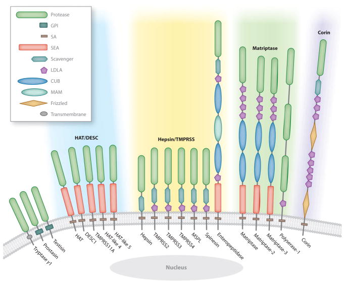
Analysis of vertebrate genome sequences at the turn of the millennium revealed that a vastly larger repertoire of enzymes execute proteolytic cleavage reactions within the pericellular and extracellular environments than was anticipated from biochemical and molecular analysis. Most unexpected was the unveiling of an entire new family of structurally unique multidomain serine proteases that are anchored directly to the plasma membrane. Unlike secreted serine proteases, which function primarily in tissue repair, immunity, and nutrient uptake, these membrane-anchored serine proteases regulate fundamental cellular and developmental processes, including tissue morphogenesis, epithelial barrier function, ion and water transport, cellular iron export, and fertilization. Here the cellular and developmental biology of this fascinating new group of proteases is reviewed. Particularly highlighted is how the study of membrane-anchored serine proteases has expanded our knowledge of the range of physiological processes that require regulated proteolysis at the cell surface.


Figures

Figure 1
Figure 1
The human complement of membrane-anchored serine proteases. The single serine protease domain of tryptase γ1, prostasin, and testisin is attached to the membrane by a C-terminal type I transmembrane domain or glycophosphatidylinositol (GPI) anchor. Type II transmembrane serine proteases are attached to the membrane by a signal anchor (SA) located close to the N terminus. The stem region between the SA and the C-terminal serine protease domain contains an assortment of domains including low-density lipoprotein receptor class A (LDLA); sea urchin sperm protein, enteropeptidase, and agrin (SEA); Cls/Clr, urchin embryonic growth factor, and bone morphogenic protein-1 (CUB); meprin, A5 antigen, and receptor protein phosphatase μ (MAM); frizzled; and group A scavenger receptor. Type II transmembrane proteases are phylogenetically divided into four subfamilies (Bugge et al. 2009, Szabo et al. 2003): (1) human airway trypsin-like (HAT)/differentially expressed in squamous cell carcinoma gene (DESC), which comprises HAT, DESC1, TMPRSS11A, HAT-like 4, and HAT-like 5 (blue shading); (2) hepsin/transmembrane protease, serine (TMPRSS), which comprises hepsin, TMPRSS2, TMPRSS3, TMPRSS4, mosaic serine protease large-form (MSPL), spinesin, and enteropeptidase ( yellow shading); (3) Matriptase, which consists of matriptase, matriptase-2, matriptase-3, and polyserase-1 ( green shading); and (4) corin, which contains only corin ( purple shading).
Figure 2
Figure 2
Membrane-anchored serine protease signaling in the surface ectoderm facilitates neural tube closure. Protease-activated receptor (PAR)-dependent Rac1 activation in the surface ectoderm through Gi/o/z is required for the fusion of the neural folds. PAR-1 is activated by thrombin. PAR-2 may be activated by prostasin, acting through matriptase, or by other locally expressed membrane-anchored serine proteases including hepsin; transmembrane protease, serine 2 (TMPRSS2); TMPRSS4; and matriptase-3. Membrane-anchored serine protease activity in surface ectoderm cells is negatively regulated by hepatocyte growth factor activator inhibitor 2 (HAI-2).
Figure 3
Figure 3
Epidermal differentiation processes controlled by the matriptase-prostasin proteolytic cascade. (a) Formation of tight junctions between granular-layer keratinocytes. (b) Cytoplasmic processing of profilaggrin to filaggrin monomers by μ-calpain and complete processing to free hygroscopic and UV light–absorbing amino acids by caspase-14 and bleomycin hydrolase. (c) Epidermal lipid synthesis by transitional cells and its deposition into the intercellular space between corneocytes as lipid lamellae. (d ) Proteolytic shedding (desquamation) of the stratum corneum by kallikrein (KLK)-mediated degradation of desmogleins in desmosomes linking corneocytes. If the four processes are independent or interrelated, and what target substrate(s) is cleaved in each process by the matriptase-prostasin cascade, are unknown.
Figure 4
Figure 4
Proteolytic regulation of epithelial sodium channel (ENaC) activity. Only one α subunit is shown, and the β subunit is omitted for simplicity. (a) The unprocessed channel has low sodium currents. (b) Furin removes an autoinhibitory disulfide-bonded loop in the α subunit during ENaC trafficking to the plasma membrane to increase the open probability. Furin also partially removes a second autoinhibitory loop in the γ subunit. (c) Full ENaC activation is achieved by complete removal of the γ subunit autoinhibitory loop by prostasin. Other membrane-anchored serine proteases may activate furin-processed ENaC in a similar manner.
Figure 5
Figure 5
Matriptase-2 regulates cellular iron export. ➀ Dietary iron and iron recycled from erythrocytes are stored in enterocytes and splenic macrophages and are released to the circulation through ferroportin. ➁ Hepcidin produced by hepatocytes binds ferroportin and targets the channel for degradation. ➂ Hepcidin gene (HAMP) expression is positively regulated by bone morphogenic protein (BMP)6, which signals through the BMP receptor (BMPR)-SMAD pathway in a hemojuvelin-dependent manner. Matriptase-2 increases cellular iron export by degrading hemojuvelin to reduce hepcidin production and thus increase ferroportin levels.

References

    1. Adham IM, Nayernia K, Engel W. Spermatozoa lacking acrosin protein show delayed fertilization. Mol Reprod Dev. 1997;46:370–76. - PubMed
    1. Alef T, Torres S, Hausser I, Metze D, Tursen U, et al. Ichthyosis, follicular atrophoderma, and hypotrichosis caused by mutations in ST14 is associated with impaired profilaggrin processing. J Investig Dermatol. 2009;129:862–69. - PubMed
    1. Altamura S, D’Alessio F, Selle B, Muckenthaler M. A novel TMPRSS6 mutation that prevents protease auto-activation causes IRIDA. Biochem J. 2010;431:363–71. - PMC - PubMed
    1. Andreasen D, Vuagniaux G, Fowler-Jaeger N, Hummler E, Rossier BC. Activation of epithelial sodium channels by mouse channel activating proteases (mCAP) expressed in Xenopus oocytes requires catalytic activity of mCAP3 and mCAP2 but not mCAP1. J Am Soc Nephrol. 2006;17:968–76. - PubMed
    1. Andrews NC. Forging a field: the golden age of iron biology. Blood. 2008;112:219–30. - PMC - PubMed

Publication types

LinkOut - more resources