. 2011 Jul 1:11:72.
doi: 10.1186/1472-6750-11-72.

T4 RNA ligase 2 truncated active site mutants: improved tools for RNA analysis

Affiliations

T4 RNA ligase 2 truncated active site mutants: improved tools for RNA analysis

Sebastien Viollet et al. BMC Biotechnol. .

Abstract

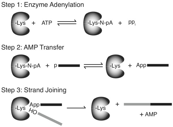
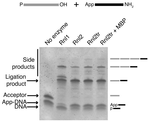
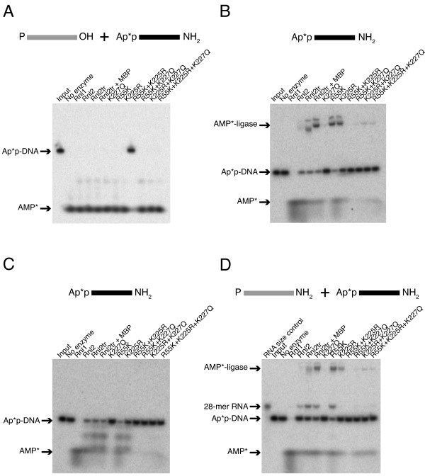
Background: T4 RNA ligases 1 and 2 are useful tools for RNA analysis. Their use upstream of RNA analyses such as high-throughput RNA sequencing and microarrays has recently increased their importance. The truncated form of T4 RNA ligase 2, comprising amino acids 1-249 (T4 Rnl2tr), is an attractive tool for attachment of adapters or labels to RNA 3'-ends. Compared to T4 RNA ligase 1, T4 Rnl2tr has a decreased ability to ligate 5'-PO4 ends in single-stranded RNA ligations, and compared to the full-length T4 Rnl2, the T4 Rnl2tr has an increased activity for joining 5'-adenylated adapters to RNA 3'-ends. The combination of these properties allows adapter attachment to RNA 3'-ends with reduced circularization and concatemerization of substrate RNA.

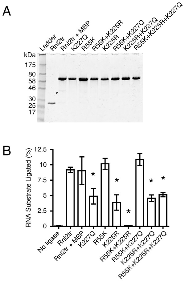
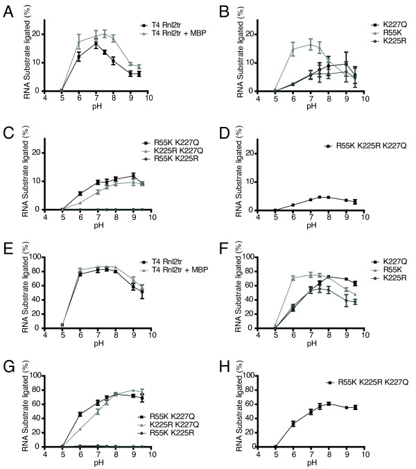
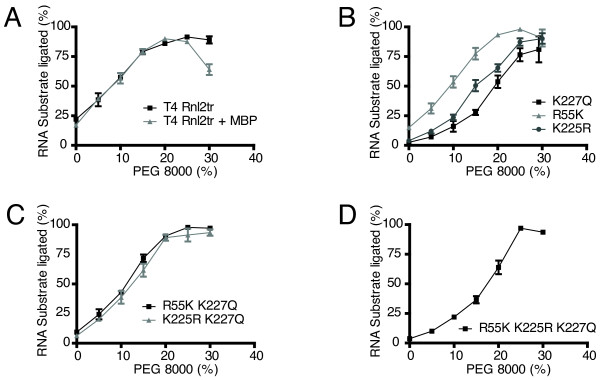
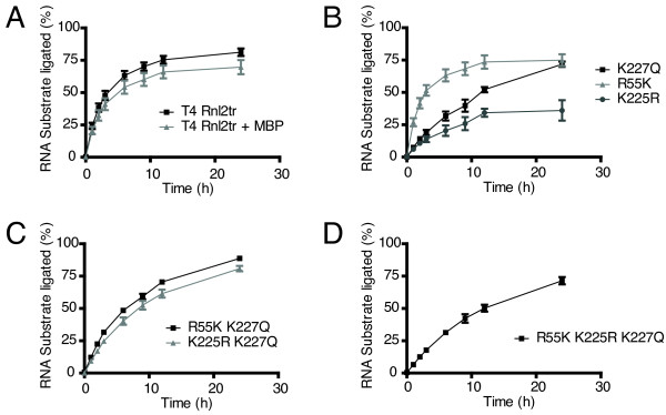
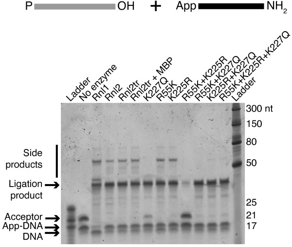
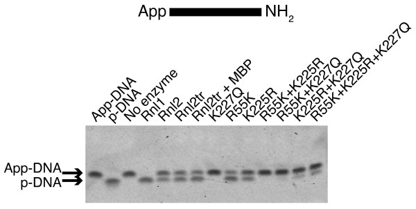
Results: With the aim of further reducing unwanted side ligation products, we substituted active site residues, known to be important for adenylyltransferase steps of the ligation reaction, in the context of T4 Rnl2tr. We characterized the variant ligases for the formation of unwanted ligation side products and for activity in the strand-joining reaction.

Conclusions: Our data demonstrate that lysine 227 is a key residue facilitating adenylyl transfer from adenylated ligation donor substrates to the ligase. This reversal of the second step of the ligation reaction correlates with the formation of unwanted ligation products. Thus, T4 Rn2tr mutants containing the K227Q mutation are useful for reducing undesired ligation products. We furthermore report optimal conditions for the use of these improved T4 Rnl2tr variants.


Figures

Figure 1
Figure 1
Schematic representation of a nucleic acid ligation reaction. In step 1, the enzyme reacts with ATP and becomes adenylated on an active site lysine residue yielding adenylated enzyme and pyrophosphate. In step 2, the AMP is transferred from the active site lysine to a 5'-phosphorylated nucleic acid donor (black). In step 3, the enzyme promotes phosphodiester bond formation between the 5'-adenylated nucleic acid donor, and a polynucleotide acceptor molecule that is 3'-hydroxylated (grey). The reaction yields a ligated polynucleotide and AMP.
Figure 2
Figure 2
Production of ligation side products by T4 RNA ligases. Intermolecular ligation reactions containing 5'-adenylated DNA adapters, 21-mer 5'-PO4 RNA acceptors and ligase (1 pmol) were incubated at 16°C overnight with 12.5% PEG 8000. Products of the reactions were resolved on denaturing 15% acrylamide gels and stained with SYBR Gold. The bands corresponding to the input nucleic acids, the DNA adapter/RNA acceptor ligation product (39 bases), and larger side products are indicated. Rnl1 = T4 RNA ligase 1, Rnl2 = T4 RNA ligase 2, Rnl2tr = T4 RNA ligase 2 truncated, Rnl2tr + MBP = T4 RNA ligase 2 truncated attached to an N-terminal maltose binding protein tag. Oligonucleotide substrates are depicted schematically above the gel. Grey lines represent RNA and black lines represent DNA.
Figure 3
Figure 3
Purification and activity of T4 RNA Ligase 2 truncated mutants. (A) Aliquots of T4 RNA ligase 2 truncated and mutants were separated on 10-20% Tris-glycine SDS polyacrylamide gels and stained with Coomassie blue. The size (in kDa) of marker polypeptides are indicated on the left. (B) Intermolecular strand-joining activity of T4 RNA ligase 2 truncated mutants under multiple turnover conditions. 10 pmol 5'-adenylated 17-mer DNA was incubated for one hour at 25°C with 5 pmol 5'- FAM-labeled 31-mer RNA. 1 pmol of each ligase was added into reaction mixture. The reaction products were resolved on denaturing 15% acrylamide gels, scanned and quantified as described in the methods section. Rnl2tr = T4 RNA ligase 2 truncated, Rnl2tr + MBP = T4 RNA ligase 2 truncated attached to an N-terminal maltose binding protein tag. All mutations indicated are substitutions in T4 Rnl2tr + MBP. Data are shown as the mean +/- SEM of at least three independent experiments. * denotes difference in means p < 0.01
Figure 4
Figure 4
Effect of pH on ligase intermolecular strand-joining activity. (A-D) Intermolecular strand-joining reactions were carried out with 10 pmol 5'-adenylated 17mer DNA, 5 pmol 31-mer 5'-FAM-labeled RNA acceptor, and ligase (1 pmol) for 1 hour at 25°C to assess the effect of pH on ligation efficiency. Ligation efficiency was determined by resolving the material in the reactions on denaturing 15% acrylamide gels and quantifying the amount of ligation product versus input nucleic acid. (E-H) Intermolecular strand-joining reactions were carried out with 10 pmol 5'-adenylated 17-mer DNA, 5 pmol 31-mer 5'-FAM-labeled RNA acceptor, and ligase (13.8 pmol) for 1 hour at 25°C to assess the effect of pH on ligation efficiency. Rnl2tr = T4 RNA ligase 2 truncated, Rnl2tr + MBP = T4 RNA ligase 2 truncated attached to an N-terminal maltose binding protein tag. All mutations indicated are substitutions in T4 Rnl2tr + MBP. Data are shown as the mean +/- SEM of at least three independent experiments.
Figure 5
Figure 5
Effect of PEG 8000 on ligase intermolecular strand-joining activity. Strand-joining reactions were carried out with 10 pmol 5'-adenylated 17-mer DNA, 5 pmol 31-mer 5'-FAM-labeled RNA acceptor, ligase (13.8 pmol), and varying amounts of PEG 8000 for 1 hour at 25°C to assess the effect of PEG on ligation efficiency. Ligation efficiency was determined by resolving the material in the reactions on denaturing 15% acrylamide gels and quantifying the amount of ligation product versus input nucleic acid. Rnl2tr = T4 RNA ligase 2 truncated, Rnl2tr + MBP = T4 RNA ligase 2 truncated attached to an N-terminal maltose binding protein tag. All mutations indicated are substitutions in T4 Rnl2tr + MBP. Data are shown as the mean +/- SEM of at least three independent experiments.
Figure 6
Figure 6
Analysis of intermolecular strand-joining over time. Strand-joining reactions were carried out with 10 pmol 5'-adenylated adapter, 5 pmol 31-mer 5'-FAM-labeled RNA acceptor, and ligase (1 pmol) over a span of 24 hours at 25°C to assess the progress of ligation reactions. Ligation efficiency was determined by resolving the material in the reactions on denaturing 15% acrylamide gels and quantifying the amount of ligation product versus input nucleic acid. Rnl2tr = T4 RNA ligase 2 truncated, Rnl2tr + MBP = T4 RNA ligase 2 truncated attached to an N-terminal maltose binding protein tag. All mutations indicated are substitutions in T4 Rnl2tr + MBP. Data are shown as the mean +/- SEM of at least three independent experiments.
Figure 7
Figure 7
Assaying the formation of side products by T4 RNA ligases. Intermolecular strand-joining reactions containing 5'-adenylated adapters, 21-mer 5'-PO4 RNA acceptors, and ligase (1 pmol) were incubated at 16°C overnight in the presence of 12.5% PEG 8000. Oligonucleotide substrates are depicted schematically above the gel. Grey lines represent RNA and black lines represent DNA. Products of the reaction were resolved on denaturing 15% acrylamide gels and stained with SYBR Gold. The bands corresponding to the input nucleic acids, the DNA adapter/RNA acceptor ligation product (39 bases), and larger side products are indicated. Ladder = size standard ladder, Rnl1 = T4 RNA ligase 1, Rnl2 = T4 RNA ligase 2, Rnl2tr = T4 RNA ligase 2 truncated, Rnl2 +MBP = T4 RNA ligase 2 truncated attached to an N-terminal maltose binding protein tag. All mutations indicated are substitutions in T4 Rnl2tr + MBP.
Figure 8
Figure 8
Deadenylation activity of T4 RNA ligase 2 truncated mutants. 5'-adenylated DNA adapters were incubated with an excess of ligase (13.8 pmol), and 12.5% PEG 8000 at 16°C overnight. Oligonucleotide substrates are depicted schematically above the gel. The contents of each sample were resolved on denaturing 15% acrylamide gels and stained with SYBR Gold to visualize nucleic acid. Deadenylation of the DNA adapter (loss of 5'-App) is indicated by a band shift of ~1 nt towards the bottom of the gel. Rnl1 = T4 RNA ligase 1, Rnl2 = T4 RNA ligase 2, Rnl2tr = T4 RNA ligase 2 truncated, Rnl2 +MBP = T4 RNA ligase 2 truncated attached to an N-terminal maltose binding protein tag. All mutations indicated are substitutions in T4 Rnl2tr + MBP.
Figure 9
Figure 9
Following AMP during ligation reactions with T4 RNA ligases. (A) 22-mer DNA adapters were 5'-adenylated with α-32P-labeled ATP (see materials and methods). Intermolecular strand-joining reactions containing 10 pmol radiolabeled DNA adapter, 5 pmol 21-mer 5'-PO4 RNA acceptor, and ligase (1 pmol) were incubated overnight at 16°C in the presence of PEG 8000. Reaction products were resolved on a denaturing 15% acrylamide gel and radioactive molecules were visualized by exposure to Phosphor screens. The resulting products were either free AMP in solution (AMP*) or the adapter remaining adenylated (Ap*p-DNA). Oligonucleotide substrates are depicted schematically above the gel. Grey lines represent RNA and black lines represent DNA. P* denotes 32P-phosphate. (B) Determining the fate of AMP upon T4 RNA ligase-dependent deadenylation. Reactions containing radiolabeled DNA adapter (10 pmol) and ligase (14 pmol) were incubated overnight at 16°C in the presence of 12.5% PEG 8000. Oligonucleotide substrates are depicted schematically above the gel. P* denotes 32P-phosphate. Reaction products were resolved and visualized as in (A). The resulting products were either free AMP in solution (AMP*), the adapter remaining adenylated (Ap*p-DNA), or AMP covalently bound to the ligase (AMP*-ligase). The lane labeled input contains only Ap*p-DNA. (C) Reactions identical to those in (B) were treated with Proteinase K prior to gel electrophoresis and detection. (D) Reactions containing 10 pmol radiolabeled DNA adapter, 5 pmol 28-mer [5'-PO4, 3'-blocked] RNA acceptor, and ligase (1 pmol) were incubated, resolved and detected as in (A). The resulting products were either free AMP in solution (AMP*), adenylated adapter (Ap*p-DNA), or Ap*p-28-mer RNA. The lane labeled RNA size control contains 5'-32PO4 RNA, and the lane labeled input contains only Ap*p-DNA. Oligonucleotide substrates are depicted schematically above the gel. Grey lines represent RNA and black lines represent DNA. P* denotes 32P-phosphate. In all panels, Rnl1 = T4 RNA ligase 1, Rnl2 = T4 RNA ligase 2, Rnl2tr = T4 RNA ligase 2 truncated, Rnl2 +MBP = T4 RNA ligase 2 truncated attached to an N-terminal maltose binding protein tag. All mutations indicated are substitutions in T4 Rnl2tr + MBP.

References

    1. Pascal JM. DNA and RNA ligases: structural variations and shared mechanisms. Curr Opin Struct Biol. 2008;18:96–105. doi: 10.1016/j.sbi.2007.12.008. - DOI - PubMed
    1. Hartmann B, Valcárcel J. Decrypting the genome's alternative messages. Curr Opin Cell Biol. 2009;21:377–386. doi: 10.1016/j.ceb.2009.02.006. - DOI - PubMed
    1. Ho CK, Shuman S. Bacteriophage T4 RNA ligase 2 (gp24.1) exemplifies a family of RNA ligases found in all phylogenetic domains. Proc Natl Acad Sci USA. 2002;99:12709–12714. doi: 10.1073/pnas.192184699. - DOI - PMC - PubMed
    1. Nandakumar J, Shuman S, Lima CD. RNA ligase structures reveal the basis for RNA specificity and conformational changes that drive ligation forward. Cell. 2006;127:71–84. doi: 10.1016/j.cell.2006.08.038. - DOI - PubMed
    1. Ho CK, Wang LK, Lima CD, Shuman S. Structure and mechanism of RNA ligase. Structure. 2004;12:327–339. - PubMed

MeSH terms

LinkOut - more resources