Comparative Study
. 2011 Jul 2;12(1):89.
doi: 10.1186/1465-9921-12-89.

Real time analysis of β(2)-adrenoceptor-mediated signaling kinetics in human primary airway smooth muscle cells reveals both ligand and dose dependent differences

Affiliations
Comparative Study

Real time analysis of β(2)-adrenoceptor-mediated signaling kinetics in human primary airway smooth muscle cells reveals both ligand and dose dependent differences

Charlotte K Billington et al. Respir Res. .

Abstract

Background: β2-adrenoceptor agonists elicit bronchodilator responses by binding to β2-adrenoceptors on airway smooth muscle (ASM). In vivo, the time between drug administration and clinically relevant bronchodilation varies significantly depending on the agonist used. Our aim was to utilise a fluorescent cyclic AMP reporter probe to study the temporal profile of β2-adrenoceptor-mediated signaling induced by isoproterenol and a range of clinically relevant agonists in human primary ASM (hASM) cells by using a modified Epac protein fused to CFP and a variant of YFP.

Methods: Cells were imaged in real time using a spinning disk confocal system which allowed rapid and direct quantification of emission ratio imaging following direct addition of β2-adrenoceptor agonists (isoproterenol, salbutamol, salmeterol, indacaterol and formoterol) into the extracellular buffer. For pharmacological comparison a radiolabeling assay for whole cell cyclic AMP formation was used.

Results: Temporal analysis revealed that in hASM cells the β2-adrenoceptor agonists studied did not vary significantly in the onset of initiation. However, once a response was initiated, significant differences were observed in the rate of this response with indacaterol and isoproterenol inducing a significantly faster response than salmeterol. Contrary to expectation, reducing the concentration of isoproterenol resulted in a significantly faster initiation of response.

Conclusions: We conclude that confocal imaging of the Epac-based probe is a powerful tool to explore β2-adrenoceptor signaling in primary cells. The ability to analyse the kinetics of clinically used β2-adrenoceptor agonists in real time and at a single cell level gives an insight into their possible kinetics once they have reached ASM cells in vivo.


Figures

Figure 1
Figure 1
Isoproterenol-induced changes in cyclic AMP activity in a single hASM cell imaged via confocal microscopy using the altered emission profile of CFP-Epac(dDEP,CD)-VENUS as a readout. CFP-Epac(dDEP,CD)-VENUS expression 48 hours post-transfection is shown in panel A (CFP excited with 440 nm and 480 nm CFP emission fluorescence recorded). Panel B shows uncorrected FRET in the same cell under basal conditions (CFP excited with 440 nm and 535 nm FRET emission fluorescence recorded). Panel C is a representative trace of the altered emission ratio (essentially a change in FRET) in real time in response to 10 μM isoproterenol (+ Iso).
Figure 2
Figure 2
Cyclic AMP activation induced by a range of doses of isoproterenol in hASM cells using (A) CFP-Epac(dDEP,CD)-VENUS activation or (B) 3H-cyclic AMP formation as a readout. (A) For Epac-based studies single hASM cells expressing CFP-Epac(dDEP,CD)-VENUS were excited at 440 nm and the emission ratio (470/535) changes collected in real time. The maximal ratio was recorded and plotted. Data are expressed as fold over basal. Each data point represents the mean (± SEM) of 3-9 separate experiments. (B) For studies investigating 3H-cyclic AMP formation, monolayers of hASM cells in 24 well plates were labelled with 3H-adenine for 2 hours and then exposed to the appropriate concentrations of isoproterenol for 5 minutes. The reaction was terminated by addition of hydrochloric acid and total 3H-labelled cyclic AMP was collected via column-based separation and quantified by scintillation counting [13]. Data are expressed as fold over basal. Each data point represents the mean (± SEM) of 3-6 experiments.
Figure 3
Figure 3
Cyclic AMP activation induced by single doses of a range of β2-adrenoceptor agonists in hASM cells using (A) CFP-Epac(dDEP,CD)-VENUS activation or (B) 3H-cyclic AMP formation as a readout. β2-adrenoceptor agonists studied were isoproterenol (10 μM), salbutamol (1 μM), salmeterol (100 nM), indacaterol (1 μM) and formoterol (1 μM). (A) For FRET-based studies single hASM cells expressing CFP-Epac(dDEP,CD)-VENUS were excited at 440 nm and the emission ratio (470/535) changes collected in real time. The maximum ratio observed was recorded and plotted. Data are expressed as fold over basal. Each data point represents the mean (± SEM) of 6-16 separate experiments. **denotes P < 0.01. (B) For studies investigating 3H-cyclic AMP formation, monolayers of hASM cells in 24 well plates were labelled with 3H-adenine for 2 hours and then exposed to the appropriate concentrations of β2-adrenoceptor agonist for 5 minutes. The reaction was terminated by addition of hydrochloric acid and total 3H-labelled cyclic AMP was collected via column-based separation and quantified by scintillation counting [13]. Data are expressed as fold over basal. Each data point represents the mean (± SEM) of 3-9 experiments. * denotes P < 0.05, **denotes P < 0.01.
Figure 4
Figure 4
Time taken for a range of β2-adrenoceptor agonists to (A) initiate an increase in CFP-Epac(dDEP,CD)-VENUS activation and (B) to induce a maximal response c.f. initiation. The time between response initiation and maximal response is shown in C. The labelled trace in D depicts how these data were obtained from real time traces. The β2-adrenoceptor agonists studied were isoproterenol (10 μM), salbutamol (1 μM), salmeterol (100 nM), indacaterol (1 μM) and formoterol (1 μM). Single hASM cells expressing CFP-Epac(dDEP,CD)-VENUS were excited at 440 nm and the emission ratio (470/535) changes collected in real time. Each data point represents the mean (± SEM) of 6-19 experiments. * denotes P < 0.05, ***denotes P < 0.001.
Figure 5
Figure 5
Re-analysis of data shown in figure 4 concentrating on the response observed in the first 30 seconds after initiation i.e. at a timepoint whereby probe saturation has not occurred. Data was analysed via (A) Area Under the Curve (AUC). B outlines the region utilised for these additional analyses. Each data point represents the mean (± SEM) of 6-19 experiments.* denotes P < 0.05, ** denotes P < 0.01.
Figure 6
Figure 6
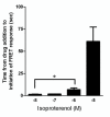
Time taken for a range of concentrations of the β2-adrenoceptor agonist isoproterenol (10-8M-10-5M) to initiate an increase in CFP-Epac(dDEP,CD)-VENUS activation. These data were obtained from real time traces comparable to those depicted in Figure 4D. Single hASM cells expressing CFP-Epac(dDEP,CD)-VENUS were excited at 440 nm and the emission ratio (470/535) changes collected in real time. Each data point represents the mean (± SEM) of 4-19 experiments. * denotes P < 0.05.

References

    1. Hall IP TA. In: Asthma. 3. Clark TH GS, Lee TH, editor. London: Chapman and Hall; 1992. Beta-agonists; pp. 341–365.
    1. Sears MR, Lotvall J. Past, present and future-beta2-adrenoceptor agonists in asthma management. Respir Med. 2005;99(2):152–170. doi: 10.1016/j.rmed.2004.07.003. - DOI - PubMed
    1. Cazzola M, Matera MG. Novel long-acting bronchodilators for COPD and asthma. Br J Pharmacol. 2008;155(3):291–299. - PMC - PubMed
    1. Billington CK, Penn RB. Signaling and regulation of G protein-coupled receptors in airway smooth muscle. Respir Res. 2003;4(1):2. doi: 10.1186/rr195. - DOI - PMC - PubMed
    1. Roscioni SS, Kistemaker LE, Menzen MH, Elzinga CR, Gosens R, Halayko AJ, Meurs H, Schmidt M. PKA and Epac cooperate to augment bradykinin-induced interleukin-8 release from human airway smooth muscle cells. Respir Res. 2009;10:88. doi: 10.1186/1465-9921-10-88. - DOI - PMC - PubMed

Publication types

MeSH terms

LinkOut - more resources