. 2011 Jul 26;21(14):1167-75.
doi: 10.1016/j.cub.2011.05.048. Epub 2011 Jun 30.

Structural organization of the actin cytoskeleton at sites of clathrin-mediated endocytosis

Affiliations

Structural organization of the actin cytoskeleton at sites of clathrin-mediated endocytosis

Agnieszka Collins et al. Curr Biol. .

Abstract

Background: The dynamic actin cytoskeleton plays an important role in clathrin-mediated endocytosis (CME). However, its exact functions remain uncertain as a result of a lack of high-resolution structural information regarding actin architecture at endocytic sites.

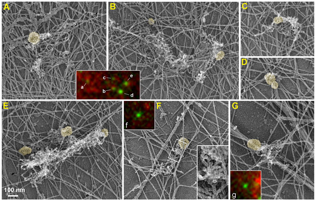
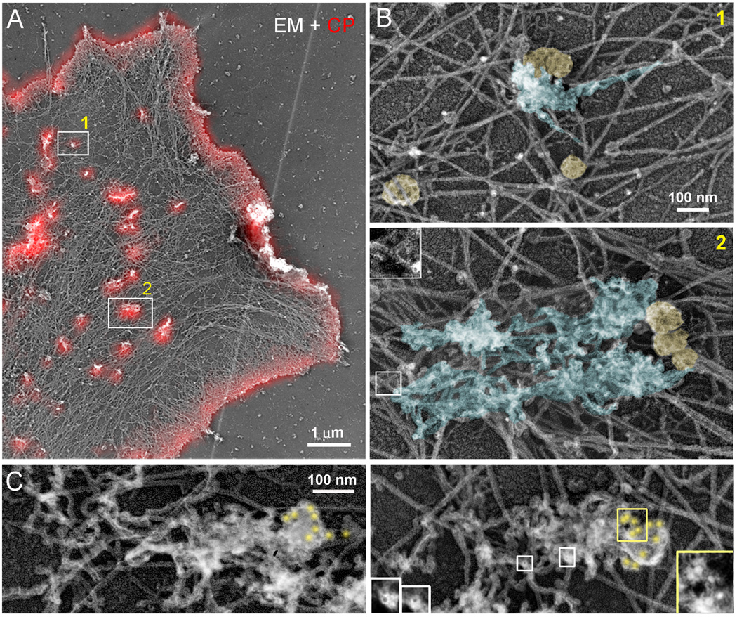
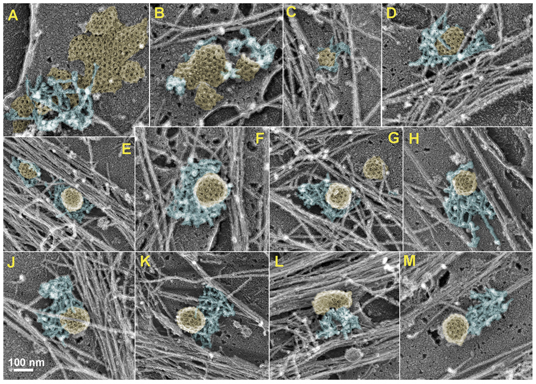
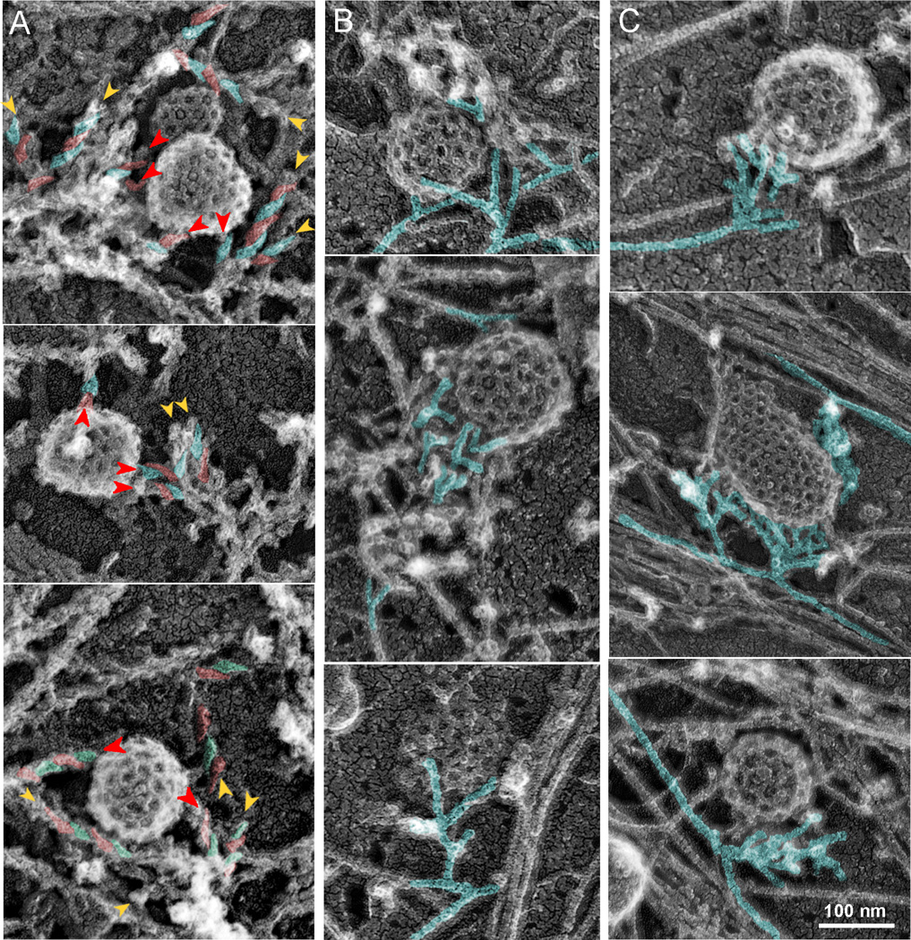
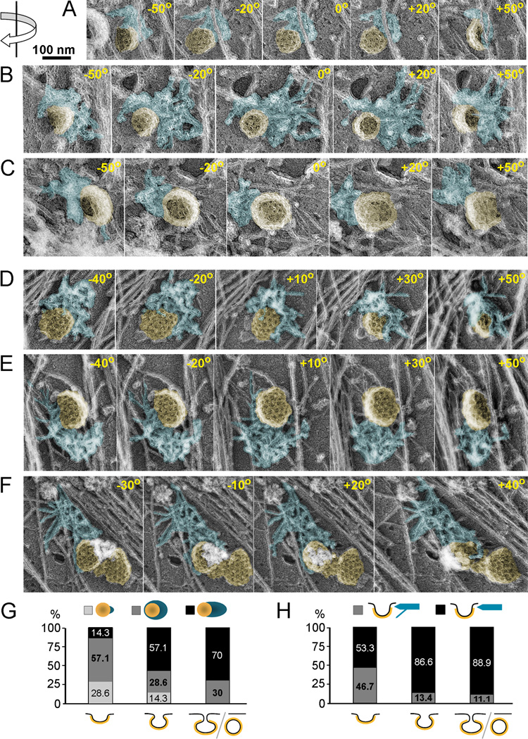
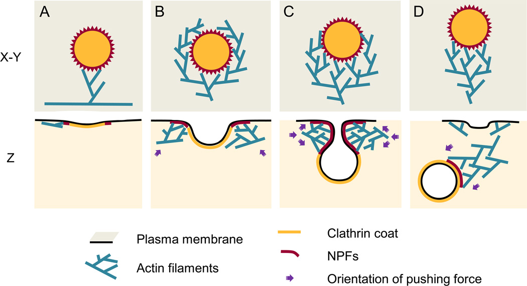
Results: Using platinum replica electron microscopy in combination with electron tomography, we found that actin patches associated with clathrin-coated structures (CCSs) in cultured mouse cells consist of a densely branched actin network, in which actin filament barbed ends are oriented toward the CCS. The shape of the actin network varied from a small lateral patch at the periphery of shallow CCSs, to a collar-like arrangement around partly invaginated CCSs with actin filament barbed ends abutting the CCS neck, to a polarized comet tail in association with highly constricted or fully endocytosed CCSs.

Conclusions: Our data suggest that the primary role of the actin cytoskeleton in CME is to constrict and elongate the bud neck and drive the endocytosed vesicles from the plasma membrane. Moreover, in these processes, barbed ends directly push onto the load, as in a conventional propulsion mechanism. Based on our findings, we propose a model for initiation, evolution, and function of the dendritic actin network at CCSs.


Figures

Figure 1
Figure 1. Correlative light and EM of GFP-CLC-expressing B16F1 cell
(A–G) EM of a detergent-extracted cell showing CCSs (yellow) that peripherally associate with ruffle-like (A–C, E) or comet-tail-like (F, G) patches of dendritic network, whereas a double CCS (D) has very little, if any, dendritic actin network. Boxed region in F is enlarged in the inset to show branched filaments. Color insets show fluorescence images of GFP-CLC (green) and phalloidin staining (red) of the CCSs labeled by corresponding letters. The CCSs labeled d, f, and g do not show distinct F-actin enrichment in their vicinity by fluorescence staining, but associate with dendritic actin networks in EM images. See Supplemental figure S1 for additional images and stereo views of CCSs.
Figure 2
Figure 2. Correlative light and EM of GFP-CPβ-expressing B16F1 cell and clathrin immunogold staining of nonexpressing cells
(A) EM of GFP-CPβ-expressing cell overlaid with GFP-CPβ fluorescence in red. (B) Enlarged EM images of boxed regions in B show dendritic comet-tails (blue) associated with CCSs (yellow). Boxed region in panel 2 is further enlarged in inset to show a branch. (C) Clathrin immunogold EM. Gold particles (yellow) accumulate at the tips of comet tails. Yellow and white boxes are enlarged in corresponding insets to show gold particles and tips of actin filaments, respectively.
Figure 3
Figure 3. CCS-associated actin patches in unroofed cells
(A–M) EM images showing clathrin plagues (A,B), shallow (C,D) and deeply invaginated (E–M) CCSs associated with dendritic actin patches of variable geometry, such as small pieces of dendritic network (A, B), collar-like (C–J), and comet tail-like (K–M) networks. See Supplemental figures S2 and S3 for fluorescence, EM and stereo EM images.
Figure 4
Figure 4. Orientation and origin of actin filaments in CCS-associated dendritic patches
EM of unroofed cells with (A) or without (B,C) S1 decoration. (A) Asymmetric “comma-shaped” units of S1 decoration revealing the filament polarity are highlighted in alternating red and cyan colors on some filaments. The head of the “comma” is oriented toward the pointed end. Barbed ends (arrowheads) face the CCS. Red arrowheads mark actin filaments terminating on CCSs. (B) The 70° angle between branched filaments (cyan) is predominantly oriented toward the CCS. (C) Dendritic actin network appears to originate from one (top and bottom) or two (middle) points on nearby long actin filaments (cyan). See Supplemental figure S3 for stereo views of CCSs.
Figure 5
Figure 5. Electron tomography of CCS-associated dendritic patches in unroofed cells
(A–F) Examples of actin patches (blue) associated with CCSs (yellow) shown as montages of selected frames from single axis tilt series acquired every 1° using FEI CM300-FEG electron microscope (A–C) or every 10° using Philips CM120 electron microscope (D–F). The tilt angles are shown in each frame. Direction of rotation is indicated by arrow at the upper left corner. Shallow (A, D), deeply invaginated (B, C, E), and constricted (F) CCSs are associated with actin patches primarily at their necks; some filaments in D hang over the CCS dome. (G, H) Correlation of three dimensional actin patch geometry with the degree of CCS invagination. (G) Actin patch geometry in the plane of the plasma membrane. Percentage of CCSs associated with a small patch (light grey), collar-shaped patch (dark grey) or comet tail (black) is plotted for each degree of invagination (shallow, deep, or constricted/endocytosed). (H) Actin patch geometry in the Z direction. Percentage of CCSs interacting with actin patch exclusively at the neck (black) or predominantly at the neck with few filaments extending over the dome (grey) are plotted for each degree of CCS invagination. Percentage numbers for each category are shown in plots (G and H). At least 11 CCSs are scored for each CCS category. See Supplemental movies 1–4 for video versions of tilt series shown in A–F.
Figure 6
Figure 6. Model for actin assembly, organization, and function during CME
Individual steps of assembly (A–D) are shown en face (X–Y) or in profile (Z). (A) Actin assembly begins when the Arp2/3 complex is activated by NPFs (red) recruited to the periphery of the CCS and binds an actin filament in the cytoplasm. (B) Actin network expands and gradually encircles the CCS through multiple rounds of dendritic nucleation guided by NPFs. The force generated by actin polymerization at this stage may drive lateral movements of the shallow CCS. (C) Dendritic actin network surrounds the neck of the deeply invaginated CCS and promotes constriction and elongation of its neck. (D) Actin network reorganizes into a comet tail and after scission drives the vesicle away from the plasma membrane.

Comment in

References

    1. Kaksonen M, Sun Y, Drubin DG. A pathway for association of receptors, adaptors, and actin during endocytic internalization. Cell. 2003;115:475–487. - PubMed
    1. Merrifield CJ, Feldman ME, Wan L, Almers W. Imaging actin and dynamin recruitment during invagination of single clathrin-coated pits. Nat Cell Biol. 2002;4:691–698. - PubMed
    1. Kaksonen M, Toret CP, Drubin DG. Harnessing actin dynamics for clathrin-mediated endocytosis. Nat Rev Mol Cell Biol. 2006;7:404–414. - PubMed
    1. Galletta BJ, Mooren OL, Cooper JA. Actin dynamics and endocytosis in yeast and mammals. Curr Opin Biotechnol. 2010;21:604–610. - PMC - PubMed
    1. Merrifield CJ, Perrais D, Zenisek D. Coupling between clathrin-coated-pit invagination, cortactin recruitment, and membrane scission observed in live cells. Cell. 2005;121:593–606. - PubMed

Publication types