. 2011 Jul 26;21(14):1176-85.
doi: 10.1016/j.cub.2011.05.049. Epub 2011 Jun 30.

Rhythmic TMS causes local entrainment of natural oscillatory signatures

Affiliations

Rhythmic TMS causes local entrainment of natural oscillatory signatures

Gregor Thut et al. Curr Biol. .

Abstract

Background: Neuronal elements underlying perception, cognition, and action exhibit distinct oscillatory phenomena, measured in humans by electro- or magnetoencephalography (EEG/MEG). So far, the correlative or causal nature of the link between brain oscillations and functions has remained elusive. A compelling demonstration of causality would primarily generate oscillatory signatures that are known to correlate with particular cognitive functions and then assess the behavioral consequences. Here, we provide the first direct evidence for causal entrainment of brain oscillations by transcranial magnetic stimulation (TMS) using concurrent EEG.

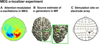
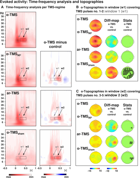
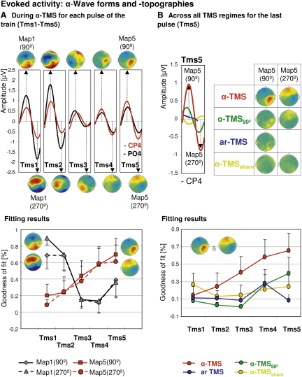
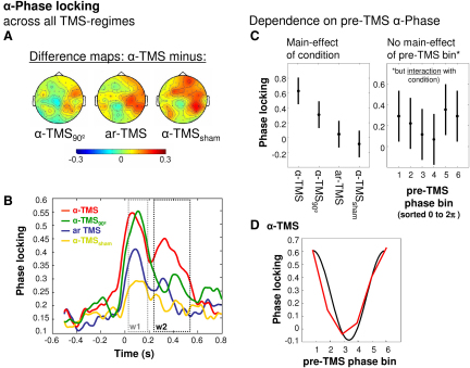
Results: We used rhythmic TMS bursts to directly interact with an MEG-identified parietal α-oscillator, activated by attention and linked to perception. With TMS bursts tuned to its preferred α-frequency (α-TMS), we confirmed the three main predictions of entrainment of a natural oscillator: (1) that α-oscillations are induced during α-TMS (reproducing an oscillatory signature of the stimulated parietal cortex), (2) that there is progressive enhancement of this α-activity (synchronizing the targeted, α-generator to the α-TMS train), and (3) that this depends on the pre-TMS phase of the background α-rhythm (entrainment of natural, ongoing α-oscillations). Control conditions testing different TMS burst profiles and TMS-EEG in a phantom head confirmed specificity of α-boosting to the case of synchronization between TMS train and neural oscillator.

Conclusions: The periodic electromagnetic force that is generated during rhythmic TMS can cause local entrainment of natural brain oscillations, emulating oscillatory signatures activated by cognitive tasks. This reveals a new mechanism of online TMS action on brain activity and can account for frequency-specific behavioral TMS effects at the level of biologically relevant rhythms.


Figures

Figure 1
Figure 1
Identification of Parietal Target Site (A) MEG grand average to the localizer task in sensor space (covert rightward minus leftward orienting of attention). (B) Estimate of most prominent α-source leading to map in (A) (right hemisphere only). The source is projected on the standard MNI brain. (C) Average position of the TMS target site projected on the electrode array (international 10–20 EEG system) that was used for EEG recordings concurrently to five-pulse TMS bursts. The TMS hot spot (coil center) was located in between CP2, CP4, P2, and P4 (closest to CP4).
Figure 2
Figure 2
Grand-Averaged Time-Frequency Plots and Topographical Analysis Comparison of α-TMS bursts (active α-TMS perpendicular to target gyrus) with all three control conditions, i.e., α-TMS90 (active α-TMS parallel to target gyrus), ar-TMS (active rapid-rate TMS in an arrhythmic regime perpendicular to target gyrus), and α-TMSsham (inactive α-TMS). (A) Time-frequency plots for electrode CP4 (closest to TMS hot spot) for all conditions (left panels) and subtractions (α-TMS minus control, right panels). w1 and w2 indicate windows of distinct early and late effects (the windows cover the entire train, which lasted 400 ms). (B) Topographies of the TMS-evoked responses for α-layer activity in the early window (w1). (C) Topographies of the TMS-evoked responses for α-layer activity in the late window (w2). The columns represent grand-average maps (left column), difference maps (α-TMS minus controls; middle columns), and corresponding t statistics (right columns). Xs indicate electrodes with statistically significant voltage differences in α-TMS relative to the corresponding control.
Figure 3
Figure 3
Evoked Activity: α-Wave Forms and Topographies (A) Top: α-waves and topographies in response to each successive single pulse during α-TMS. Waveforms are shown for electrodes CP4 (red) and PO4 (black). Map topographies are shown for the first and second part of the α-cycle (at 90° and 270° phase angle). Bottom: result of the spatial fitting procedure (spatial map correlations) for statistical evaluation of the time course of the initial (Map 190°&270°) and end maps (Map 590°&270°). (B) Top: α-waves and topographies to the last pulse of the train (Tms5) across all conditions. Bottom: result of the spatial fitting procedure (spatial map correlations) for statistical evaluation of the end α (entrainment) maps (Map 590°&270°) across pulses (Tms1–5) per condition (α-TMS, α-TMS90, ar-TMS, and α-TMSsham). Error bars represent standard error of the mean.
Figure 4
Figure 4
α-Phase Locking and Dependence on Pre-TMS Phase (A) Topographies of α-phase locking differences in window 2, expressed as change relative to baseline (left, α-TMS minus α-TMS90; middle, α-TMS minus ar-TMS; right, α-TMS minus α-TMSsham). (B) Time course of relative change of phase locking at significant electrodes with respect to baseline for all four conditions. (C) Results of two-way ANOVA on end α-phase locking (in window 2). Left: population marginal means for factor condition. Right: population marginal means for factor pre-TMS phase bin (sorted from 0 to 2π). Error bars represent 95% confidence intervals. (D) α-TMS-specific dependence of α-phase locking (w2) on pre-TMS phase bins. The black curve represents a perfect cosine function.

References

    1. Thut G., Miniussi C. New insights into rhythmic brain activity from TMS-EEG studies. Trends Cogn. Sci. (Regul. Ed.) 2009;13:182–189. - PubMed
    1. Siebner H.R., Ziemann U. Rippling the cortex with high-frequency (>100 Hz) alternating current stimulation. J. Physiol. 2010;588:4851–4852. - PMC - PubMed
    1. Klimesch W., Sauseng P., Gerloff C. Enhancing cognitive performance with repetitive transcranial magnetic stimulation at human individual alpha frequency. Eur. J. Neurosci. 2003;17:1129–1133. - PubMed
    1. Sauseng P., Klimesch W., Heise K.F., Gruber W.R., Holz E., Karim A.A., Glennon M., Gerloff C., Birbaumer N., Hummel F.C. Brain oscillatory substrates of visual short-term memory capacity. Curr. Biol. 2009;19:1846–1852. - PubMed
    1. Romei V., Gross J., Thut G. On the role of prestimulus alpha rhythms over occipito-parietal areas in visual input regulation: Correlation or causation? J. Neurosci. 2010;30:8692–8697. - PMC - PubMed

Publication types