. 2011 Jul 6;101(1):33-42.
doi: 10.1016/j.bpj.2011.05.029.

Quantification of collagen organization in the peripheral human cornea at micron-scale resolution

Affiliations

Quantification of collagen organization in the peripheral human cornea at micron-scale resolution

Craig Boote et al. Biophys J. .

Abstract

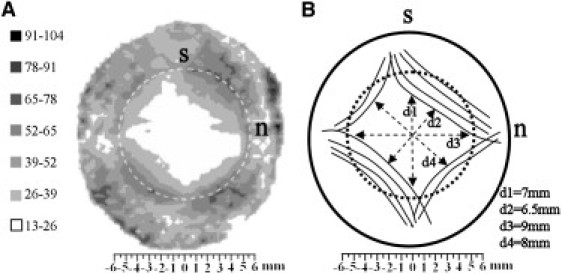
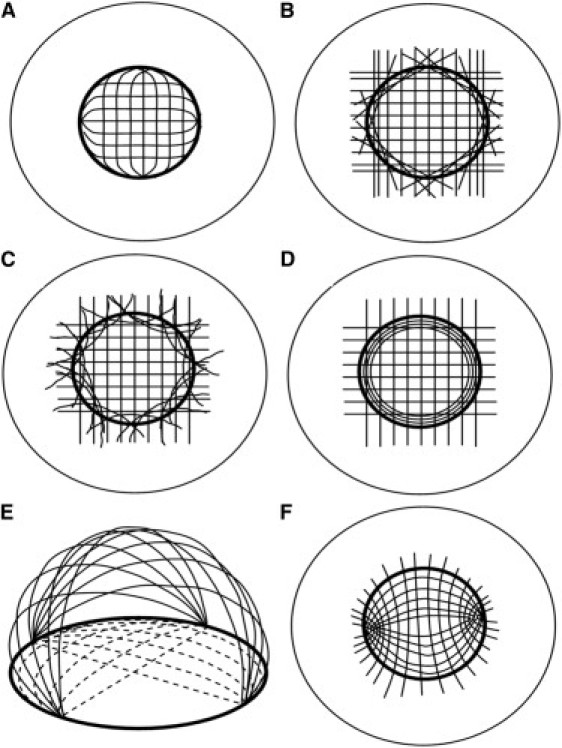
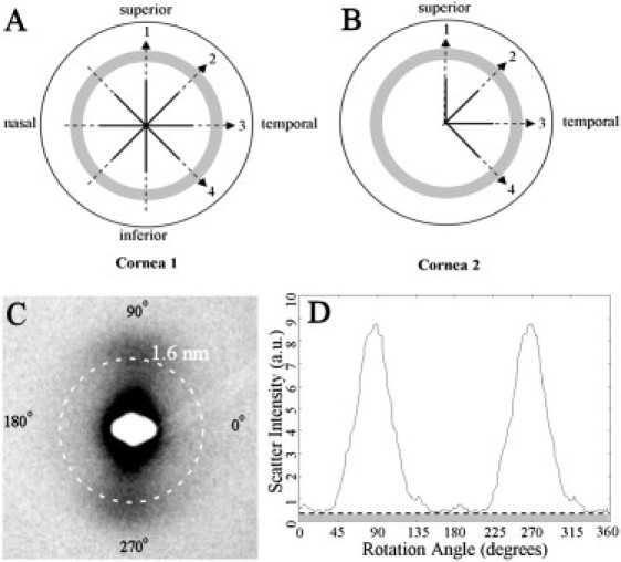
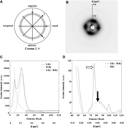
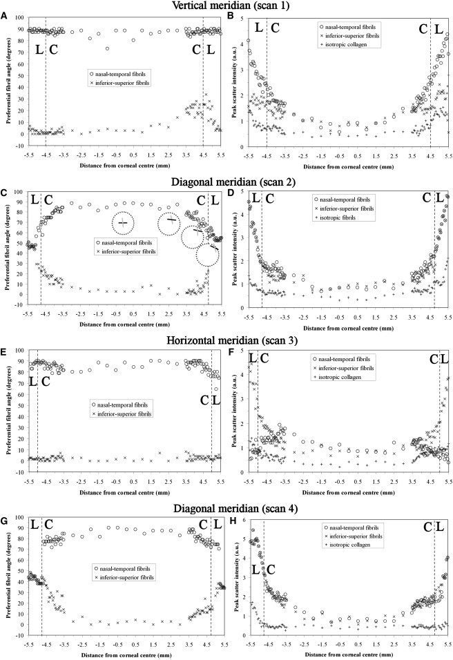
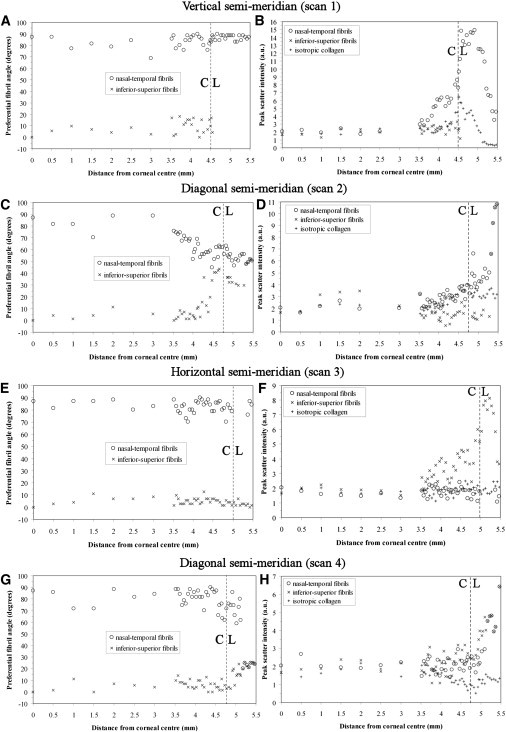
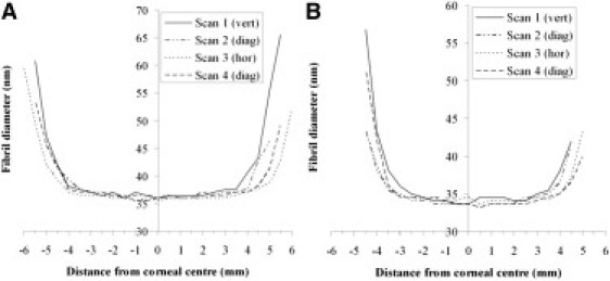
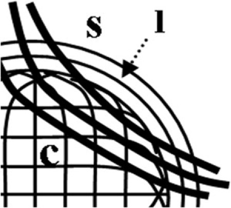
The collagen microstructure of the peripheral cornea is important in stabilizing corneal curvature and refractive status. However, the manner in which the predominantly orthogonal collagen fibrils of the central cornea integrate with the circumferential limbal collagen is unknown. We used microfocus wide-angle x-ray scattering to quantify the relative proportion and orientation of collagen fibrils over the human corneolimbal interface at intervals of 50 μm. Orthogonal fibrils changed direction 1-1.5 mm before the limbus to integrate with the circumferential limbal fibrils. Outside the central 6 mm, additional preferentially aligned collagen was found to reinforce the cornea and limbus. The manner of integration and degree of reinforcement varied significantly depending on the direction along which the limbus was approached. We also employed small-angle x-ray scattering to measure the average collagen fibril diameter from central cornea to limbus at 0.5 mm intervals. Fibril diameter was constant across the central 6 mm. More peripherally, fibril diameter increased, indicative of a merging of corneal and scleral collagen. The point of increase varied with direction, consistent with a scheme in which the oblique corneal periphery is reinforced by chords of scleral collagen. The results have implications for the cornea's biomechanical response to ocular surgeries involving peripheral incision.


Figures

Figure 1
Figure 1
(A) Contour map of aligned collagen x-ray scatter (a.u.) from a right human cornea. Superior, s, and nasal, n, positions are marked. Broken line denotes the limbus. Note the skewed diamond shape of the scatter contours, which displays mirror symmetry between the left and right eyes. (B) Proposed model of collagen fibril arrangement to explain the shape of the aligned scatter contours. The peripheral, oblique cornea is reinforced by chords of anchoring collagen of scleral origin. Figure modified from Boote et al. (13).
Figure 2
Figure 2
Previous models to explain the integration of corneal and limbal fibrils, based on data from (AE) x-ray scattering and (F) circular polarization biomicroscopy. (A) The orthogonal fibrils change direction to form the limbal annulus. (B) A discrete population of straight, tangential scleral fibrils forms the pseudoannulus. (C) Discrete, curved scleral fibrils form the limbal annulus. (D) The limbal annulus is a separate population of circular fibrils. (E) Linear belts of collagen (solid lines) run from limbus to limbus, leading to a two-dimensional projection view (broken lines) characterized by central orthogonal and peripheral annular fibrils. (F) Confocal elliptic/hyperbolic model in which fibrils loop around nasal and temporal foci. Redrawn from (AD) Meek and Boote (21), (E) Pinsky et al. (23), and (F) Misson (24).
Figure 3
Figure 3
(A and B) Scan lines on the two left human corneoscleral buttons used for microfocus WAXS. Solid line: coarse sampling (0.5 mm); broken line: fine sampling (0.05 mm). Shaded region denotes the limbus. (C) WAXS pattern from the limbal region of Cornea 1, showing the collagen intermolecular WAXS reflection centered at 1.6 nm. (D) Normalized x-ray scatter from fibrillar collagen as a function of rotation angle. Each of the 256 values in the distribution is extracted via a radial integration of the (background-subtracted) collagen WAXS peak. The clear region corresponds to preferentially aligned collagen, whereas the shaded region corresponds to isotropic collagen.
Figure 4
Figure 4
(A) Scan lines on the two right human corneoscleral buttons used for SAXS. The sampling interval was 0.5 mm and the shaded region denotes the limbus. (B) SAXS pattern from the center of Cornea 3. (C) Vertical intensity profile, I(K), through pattern shown in B. The data is folded about the pattern center. A background function, B(K), is subtracted. The collagen interference function peak arising from the short-range lateral order of the stromal fibrils can be clearly seen. The region bounded by the rectangular box is shown expanded in D. (D) A fibril transform function, F(K), is fitted to the background-subtracted data and the peak position (solid arrow) calibrated to determine the average collagen fibril diameter. The sharp third order collagen meridional peak (empty arrow) is visible merged into the equatorial pattern, and may be ignored in fitting the fibril transform.
Figure 5
Figure 5
(A, C, E, and G) Preferential angle of (centrally) i-s and n-t aligned fibril populations, as a function of distance from corneal center for Cornea 1. (B, D, F, and H) Peak x-ray scatter for aligned and isotropic fibril populations. The corneal, C, and limbal, L, regions are indicated. Insets in C depict line plots representing the local direction of n-t (black line) and i-s (gray line) populations at four selected points along diagonal scan 2.
Figure 6
Figure 6
(A, C, E, and G) Preferential angle of aligned fibril populations for Cornea 2. (B, D, F, and H) Peak x-ray scatter for aligned and isotropic fibril populations. The corneal, C, and limbal, L, regions are indicated.
Figure 7
Figure 7
Average collagen fibril diameter as a function of distance from the corneal center for (A) Cornea 3 and (B) Cornea 4.
Figure 8
Figure 8
Schematic showing possible integration of the corneal, c, and limbal, l, collagen fibrils in the right human eye, based on the current x-ray data (superonasal quadrant shown). Centrally orthogonal fibrils change direction in the peripheral cornea to fuse with the tangential fibrils of the highly reinforced limbal annulus. Chords of larger anchoring fibrils, originating in the sclera, s, cross the oblique peripheral cornea.

References

    1. Maurice D.M. The structure and transparency of the cornea. J. Physiol. 1957;136:263–286. - PMC - PubMed
    1. Komai Y., Ushiki T. The three-dimensional organization of collagen fibrils in the human cornea and sclera. Invest. Ophthalmol. Vis. Sci. 1991;32:2244–2258. - PubMed
    1. Fatt I., Weissman B. Butterworth-Heinmann; Boston, MA: 1992. Physiology of the Eye: An Introduction to the Vegetative Functions.
    1. Meek K.M. Corneal collagen—its role in maintaining corneal shape and transparency. Biophys. Rev. 2009;1:83–93. - PMC - PubMed
    1. Meek K.M., Boote C. The use of x-ray scattering techniques to quantify the orientation and distribution of collagen in the corneal stroma. Prog. Retin. Eye Res. 2009;28:369–392. - PubMed

Publication types

LinkOut - more resources