. 2011 Oct;39(18):8213-22.
doi: 10.1093/nar/gkr500. Epub 2011 Jun 30.

Structural basis for the cooperative DNA recognition by Smad4 MH1 dimers

Affiliations

Structural basis for the cooperative DNA recognition by Smad4 MH1 dimers

Nithya Baburajendran et al. Nucleic Acids Res. 2011 Oct.

Abstract

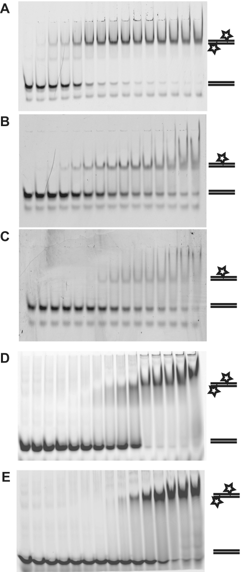
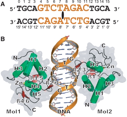
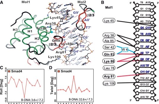
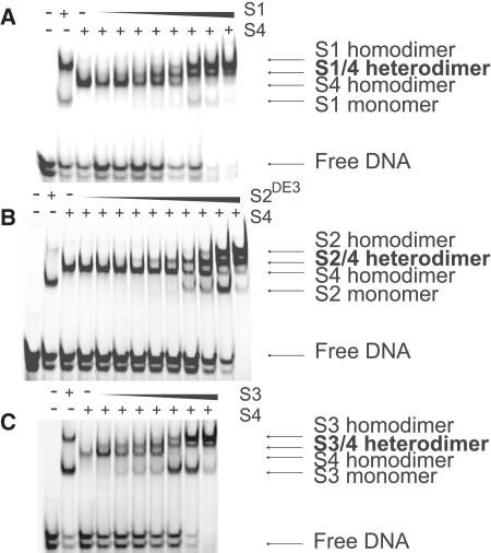
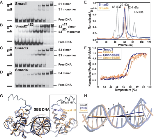
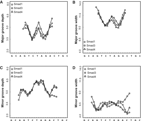
Smad proteins form multimeric complexes consisting of the 'common partner' Smad4 and receptor regulated R-Smads on clustered DNA binding sites. Deciphering how pathway specific Smad complexes multimerize on DNA to regulate gene expression is critical for a better understanding of the cis-regulatory logic of TGF-β and BMP signaling. To this end, we solved the crystal structure of the dimeric Smad4 MH1 domain bound to a palindromic Smad binding element. Surprisingly, the Smad4 MH1 forms a constitutive dimer on the SBE DNA without exhibiting any direct protein-protein interactions suggesting a DNA mediated indirect readout mechanism. However, the R-Smads Smad1, Smad2 and Smad3 homodimerize with substantially decreased efficiency despite pronounced structural similarities to Smad4. Therefore, intricate variations in the DNA structure induced by different Smads and/or variant energetic profiles likely contribute to their propensity to dimerize on DNA. Indeed, competitive binding assays revealed that the Smad4/R-Smad heterodimers predominate under equilibrium conditions while R-Smad homodimers are least favored. Together, we present the structural basis for DNA recognition by Smad4 and demonstrate that Smad4 constitutively homo- and heterodimerizes on DNA in contrast to its R-Smad partner proteins by a mechanism independent of direct protein contacts.


Figures

Figure 1.
Figure 1.
Smad4 forms a constitutive dimer on the palindromic SBE and poorly resolved monomers on other elements. Smad4 MH1 binding to (A) the palindromic SBE (TCAGTCTAGACATAC) (B) the single SBE (AGTATGTCTCAGATGA) (C) the ‘GC-BRE’ type element (CGCCTGGCGCCAGAGA) was analyzed by EMSA using 10% gels and 1 nM cy5 labeled DNA probes. Experiments were performed in triplicates. Smad4 MH1 binding to (D) the JunB promoter element with GTCT direct repeat (GACAGTCTGTCTGCC) and (E) the OPN1 promoter element with divergent palindromic GTCT repeats separated by a 1-nt spacer (TGGAGACTGTCTGGA) were analyzed by EMSA using 10% gels and 500 nM cy5-labeled DNA probes. Protein concentrations increase in a 2-fold manner to 2000 nM from left to right.
Figure 2.
Figure 2.
Overall structure of the Smad4 MH1/SBE complex. (A) Sequence of the 16-bp SBE palindromic DNA used for crystallization. The core SBE palindrome GTCTAGAC is shown in orange. (B) The overall structure of the dimeric Smad1 MH1 bound to SBE DNA shown as cartoon and semi-transparent van-der-Waals surface. α-helices are colored in green, β-sheets in red and loop regions in black.
Figure 3.
Figure 3.
DNA recognition by Smad4. (A) Protein–DNA interactions of the Smad4 MH1. Arg81, Gln83 and Lys88 specifically interacting with A8, G9 and G4′ as well as residues engaged in backbone contacts are shown as ball-and-sticks. The water molecule mediating interactions with Ser42 is shown as a blue sphere. Phosphate interactions are indicated with black dashes. (B) Schematic drawing of Smad4 MH1 SBE DNA interaction. Amino acids engaging in specific DNA contacts (red lines) as well as phosphate backbone contacts (black lines) and water mediated contacts (blue line) are shown. Only contacts by molecule 1 are shown for clarity. Smad4 induces deviation from the B-DNA topology. (C) Roll and (D) twist angles determined using Curves+ plotted against the SBE DNA sequences highlighting the negative roll and the over-twisting at the palindromic center. Parameters for standard B-DNA are shown and (+/–) indicates standard deviations (38). The overall topologies at the palindromic centers are similar for Smad1, Smad3 and Smad4 bound DNA and details are given in Supplementary Figures S3 and S4.
Figure 4.
Figure 4.
Heteromerization between Smad4 and R-Smads. An amount of 600 nM of Smad4 MH1 was pre-incubated with 2000 nM SBE DNA and 2-fold serial dilutions (right to left) of the (A) Smad1 MH1 (S1, 1600 nM in lane 1 and 2-fold serial dilution starting from 6400 nM), (B) the Smad2ΔE3 MH1 (S2, 320 nM in lane 1 and 2-fold serial dilution starting from 2560 nM) and (C) the Smad3 MH1 (S3, 800 nM in lane 1 and 2-fold serial dilution starting from 6400 nM) were added and the reactions were analyzed by EMSAs. The various DNA bound Smad complexes are marked. The titrations reveal that R-Smad/Smad4 heterodimers form at lower concentrations than R-Smad mono- or homodimers indicating that heterodimerization is the preferred binding mode on the palindromic SBE.
Figure 5.
Figure 5.
Comparison of Smad1, Smad3 and Smad4. EMSAs to compare the binding of (A) the Smad1 MH1, (B) the Smad2ΔE3 MH1, (C) the Smad3 MH1 and (D) the Smad4 MH1 to 2.5 μM SBE DNA reveal substantially different homodimerization patterns. (E) Gel filtration chromatograms showing that the Smad3 MH1 and the Smad4 MH1 elute with overlapping peaks. The chromatogram of a molecular weight standard is shown in grey (22,27). (F) Melting curves for the Smad3 and Smad4 MH1 in the absence and presence of DNA were recorded employing circular dichroism spectroscopy as described (22). The data indicate that the Smad4 MH1 is structurally stabilized upon DNA binding whereas the Smad3 MH1 is not. (G) Superposition of the MH1/DNA structures for Smad1, Smad3 and Smad4 emphasizing overall structural similarities with the notable exceptions of the ‘open’ conformation of helix 1 of Smad1 and an N-terminally shortened helix 2 seen for Smad4. (H) Overlay of the helical axes calculated with Curves+ and cartoons of the SBE DNAs bound to Smad1 (black), Smad3 (blue) and Smad4 MH1 (orange) illustrating differences of the overall curvature of the double helix.
Figure 6.
Figure 6.
Major groove depth (A) and width (B) as well as minor groove depth (C) and width (D) were calculated using Curves+ for Smad1 MH1 (circles), Smad3 MH1 (squares) and Smad4 MH1 (triangles) bound SBE DNA. See Supplementary Table S3 for Pearson’s product moment correlation analysis of helical parameters.

References

    1. Whitman M. Smads and early developmental signaling by the TGFbeta superfamily. Genes Dev. 1998;12:2445–2462. - PubMed
    1. Massagué J. TGF-beta signal transduction. Annu. Rev. Biochem. 1998;67:753–791. - PubMed
    1. Hoodless P, Haerry T, Abdollah S, Stapleton M, O’Connor M, Attisano L, Wrana J. MADR1, a MAD-related protein that functions in BMP2 signaling pathways. Cell. 1996;85:489–500. - PubMed
    1. Kawai S, Faucheu C, Gallea S, Spinella-Jaegle S, Atfi A, Baron R, Roman S. Mouse smad8 phosphorylation downstream of BMP receptors ALK-2, ALK-3, and ALK-6 induces its association with Smad4 and transcriptional activity. Biochem. Biophys. Res. Commun. 2000;271:682–687. - PubMed
    1. Macías-Silva M, Abdollah S, Hoodless P, Pirone R, Attisano L, Wrana J. MADR2 is a substrate of the TGFbeta receptor and its phosphorylation is required for nuclear accumulation and signaling. Cell. 1996;87:1215–1224. - PubMed

Publication types

Associated data