. 2011 Oct;39(18):8122-34.
doi: 10.1093/nar/gkr521. Epub 2011 Jun 30.

A multifactor regulatory circuit involving H-NS, VirF and an antisense RNA modulates transcription of the virulence gene icsA of Shigella flexneri

Affiliations

A multifactor regulatory circuit involving H-NS, VirF and an antisense RNA modulates transcription of the virulence gene icsA of Shigella flexneri

Chi Nhan Tran et al. Nucleic Acids Res. 2011 Oct.

Abstract

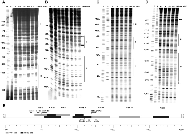
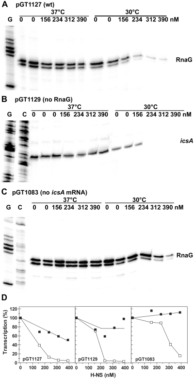
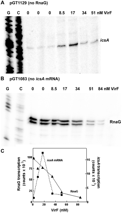
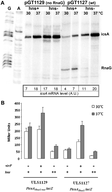
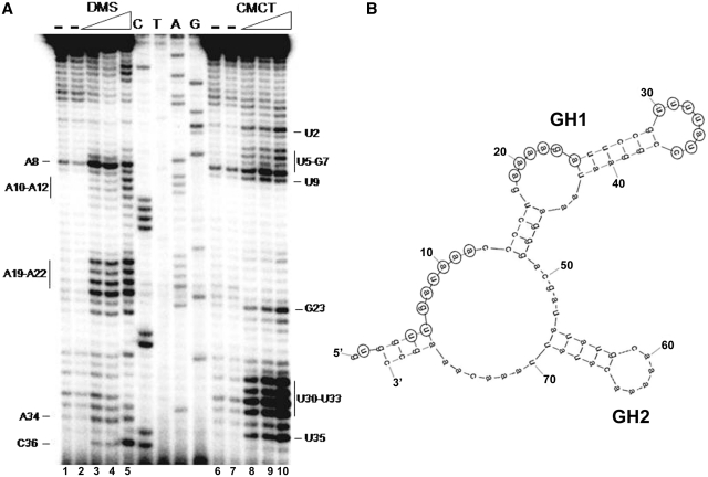
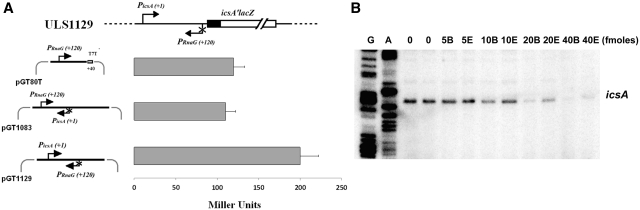
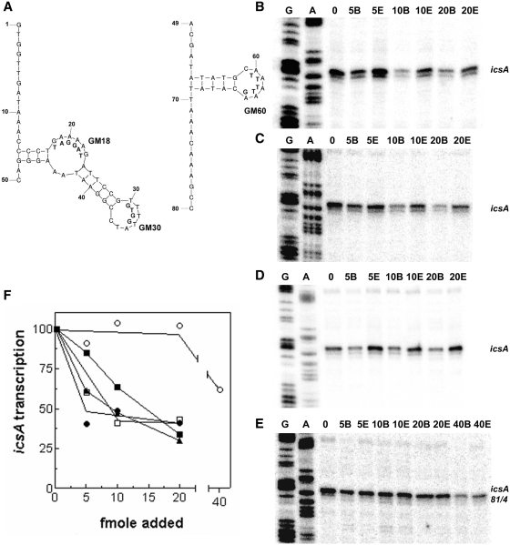
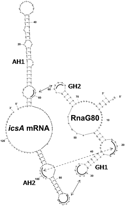
The icsA gene of Shigella encodes a structural protein involved in colonization of the intestinal mucosa by bacteria. This gene is expressed upon invasion of the host and is controlled by a complex regulatory circuit involving the nucleoid protein H-NS, the AraC-like transcriptional activator VirF, and a 450 nt antisense RNA (RnaG) acting as transcriptional attenuator. We investigated on the interplay of these factors at the molecular level. DNase I footprints reveal that both H-NS and VirF bind to a region including the icsA and RnaG promoters. H-NS is shown to repress icsA transcription at 30°C but not at 37°C, suggesting a significant involvement of this protein in the temperature-regulated expression of icsA. We also demonstrate that VirF directly stimulates icsA transcription and is able to alleviate H-NS repression in vitro. According to these results, icsA expression is derepressed in hns- background and overexpressed when VirF is provided in trans. Moreover, we find that RnaG-mediated transcription attenuation depends on 80 nt at its 5'-end, a stretch carrying the antisense region. Bases engaged in the initial contact leading to sense-antisense pairing have been identified using synthetic RNA and DNA oligonucleotides designed to rebuild and mutagenize the two stem-loop motifs of the antisense region.


Figures

Figure 1.
Figure 1.
H-NS and VirF show multiple binding sites on icsA regulatory region. Supercoiled plasmids pKG673 (A, C and D) and pKG450 (B), containing the promoter and part of the coding region of icsA, were incubated at the indicated concentrations of either H-NS (A and B) or VirF (C and D) expressed as dimer and monomer, respectively. Samples were processed as described in ‘Materials and methods’ section, using the oligos GBA (A), ACC9 (B), G–100 (C) and G+370 E (D) as primers. Lanes G, A and C represent the sequencing reactions using the same primers. Protections and sites hypersensitive to DNase I are indicated by vertical lines and arrows, respectively. The diagram shows the icsA promoter region from positions −100 to +390 (E). H-NS and VirF DNase I protections, −10 and −35 consensus elements, transcriptional start points of icsA and RnaG (ts) and the icsA initiation triplet (ATG) are indicated.
Figure 2.
Figure 2.
H-NS inhibits icsA and RnaG transcription. In vitro transcription was carried out at 30 and 37°C on ~200 ng of the supercoiled plasmid templates pGT1127 (A), pGT1129 (B) and pGT1083 (C) in the presence of the indicated concentrations of H-NS. The icsA and RnaG transcripts were detected by primer extension using the oligos G+110 and G+50, respectively, and quantified by imager (D). The transcription levels were normalized taking as 100%, the basal activity of the promoter measured at 30 and 37°C, in absence of H-NS. Values of the duplicated and/or tripled ‘0’ lanes were averaged and such value taken as 100%. All points of each individual set of samples were expressed as percentage of the corresponding ‘0’ samples. Lanes G and C represent the sequencing reactions using the same primer.
Figure 3.
Figure 3.
VirF stimulates icsA and represses RnaG transcription. In vitro transcription was carried out for 20 min at 37°C on the supercoiled plasmid templates pGT1129 (A) and pGT1083 (B) in presence of the indicated concentrations of VirF. The icsA and RnaG transcripts were detected essentially as described in Figure 2. Lanes G and C represent the sequencing reactions using the same primer. Radioactivity associated with icsA and RnaG transcripts is plotted versus VirF concentration (C).
Figure 4.
Figure 4.
Effect of H-NS and VirF on the in vivo expression of icsA. (A) Escherichia coli wt (HMG11) and its isogenic hns- strain (HMG9) harboring pGT1127 or pGT1129 plasmids were grown at 30 and 37°C in LB broth to A600 = 0.5. Aliquots of each culture were processed for total RNA extraction and 10 µg of each sample were analyzed by primer extension using the oligos G+50 and G+110 to detect RnaG and icsA mRNA, respectively. (B) Esherichia coli strains ULS1127 and ULS1129, carrying a wt hns gene (indicated with hns+) and a single-copy chromosomal PicsA-lacZ fusion with, respectively, a functional RnaG promoter (PicsA/RnaG-lacZ) or a defective one (PicsA/RnaGmut-lacZ), were used to assay β-galactosidase activity in the presence of pMYSH6504 (a plasmid harboring the S. flexneri virF gene indicated with virF+) or pDIA510 (a plasmid carrying extracopies of the E. coli hns gene indicated with hns++). Strains were grown in LB broth at 30 or 37°C to A600 = 0.4. The values represent the average of at least three independent experiments and the standard deviation is indicated.
Figure 5.
Figure 5.
Secondary structure at 5′-end of RnaG. (A) Chemical probing of RnaG80 (nucleotides 1–80). Purified RnaG80 (2 pmol) was treated with increasing amounts of the single-strand specific reagents dimethyl sulfate (DMS) (0%, lanes 1 and 2; 0.22%, lane 3; 0.46%, lanes 4; 0.78%, lane 5) and 1-cyclohexyl-3-(2-morpholinoethyl) carbodiimide metho-p-toluene sulfonate (CMCT) (0 mg/ml, lanes 6 and 7; 1.5 mg/ml, lane 8; 3 mg/ml, lane 9; 6 mg/ml, lane 10) as described in ‘Materials and Methods’ section. Modified nucleotides were detected by primer extension using the oligo G+40H and accessible sites were evaluated comparing samples incubated in the absence (−) and in the presence of either DMS or CMCT. Lanes C, T, A and G correspond to DNA sequencing ladders made with the same primer. (B) Schematic representation of the secondary structure of RnaG80. Numbering is according to 5′-end of the antisense RNA corresponding to position +120 on icsA sequence. Nucleotides reactive to DMS or CMCT in Panel A are circled. Chemical probing data were superimposed on a computer prediction generated by the MFOLD program (47).
Figure 6.
Figure 6.
RnaG80 is able to repress icsA expression. (A) The expression of a PicsA-lacZ fusion carried by E. coli strain ULS1129 was monitored in cells transformed with pGT80T, pGT1083 or pGT1129 as control. The data (±SD) represent the average of three independent experiments. (B) In vitro transcription was carried out at 37°C using 200 ng of supercoiled plasmid pGT1129 as template. Increasing amounts of RnaG80 were added to the transcription mixture either at the beginning of the reaction (prior to RNA polymerase addition, samples B) or at the end (immediately before RNA precipitation, samples E). The in vitro transcribed icsA mRNA was primer-extended with the oligo G+110.
Figure 7.
Figure 7.
Effects on icsA transcription of synthetic RNA and DNA oligonucleotides reproducing the structural motifs of RnaG. Secondary structure of DNA oligos used to construct the GH1 (left) and GH2 (right) stem–loops of RnaG (A). Mutations carried by oligos GM18, GM30 and GM60 are reported below the native sequence. In vitro transcription of icsA gene was performed, as described in the legend of Figure 6, using as templates the pGT1129 (B, C and D) and the pGT1129M (E) which synthesizes the mutated icsA81/4 mRNA (17). The increasing amounts of RNA oligo pair GR4–48/GR49–80 (B) and DNA oligo pairs G1–50/G49–80 (C and E) and G14–48/G49–80 (D) are indicated. After quantization, the radioactivity associated with icsA mRNA is expressed as ratio sample B/sample E and values are plotted as percentage assuming 100% the transcription of DNA template without oligos (F). Data from Figure 6B and from panels B, C, D and E are indicated with closed squares, open squares, closed triangles, closed circles and open circles, respectively.
Figure 8.
Figure 8.
Identification of stem–loop motifs involved in the formation of the kissing complex between RnaG and icsA mRNA. Schematic representation of the three regions (arrows) providing the initial nucleation points for the pairing of RnaG80 and icsA mRNA. Interactions occur between GH2 (positions 60–63) and AH1 (positions 59–62), as well as between GH1 (positions 18–23 and 30–36) and AH2 (positions 98–103 and 85–91).

References

    1. Sansonetti PJ. Shigellosis: an old disease in new clothes? PLoS Med. 2006;3:e354. - PMC - PubMed
    1. Prosseda G, Falconi M, Nicoletti M, Casalino M, Micheli G, Colonna B. Histone-like proteins and the Shigella invasivity regulon. Res. Microbiol. 2002;153:461–468. - PubMed
    1. Parsot C. Shigella spp. and enteroinvasive Escherichia coli pathogenicity factors. FEMS Microbiol. Lett. 2005;252:11–18. - PubMed
    1. Bernardini ML, Mounier J, d'Hauteville H, Coquis-Rondon M, Sansonetti PJ. Identification of icsA, a plasmid locus of Shigella flexneri that governs bacterial intra- and intercellular spread through interaction with F-actin. Proc. Natl Acad. Sci. USA. 1989;10:3867–3871. - PMC - PubMed
    1. Brandon LD, Goehring N, Janakiraman A, Yan AW, Wu T, Beckwith J, Goldberg MB. IcsA, a polarly localized autotransporter with an atypical signal peptide, uses the Sec apparatus for secretion, although the Sec apparatus is circumferentially distributed. Mol. Microbiol. 2003;50:45–60. - PubMed

Publication types

MeSH terms