Review
. 2011 Jul 1;25(13):1345-58.
doi: 10.1101/gad.2057811.

The diverse functions of Dot1 and H3K79 methylation

Affiliations
Review

The diverse functions of Dot1 and H3K79 methylation

Anh Tram Nguyen et al. Genes Dev. .

Abstract

DOT1 (disruptor of telomeric silencing; also called Kmt4) was initially discovered in budding yeast in a genetic screen for genes whose deletion confers defects in telomeric silencing. Since the discovery ∼10 years ago that Dot1 and its mammalian homolog, DOT1L (DOT1-Like), possess histone methyltransferase activity toward histone H3 Lys 79, great progress has been made in characterizing their enzymatic activities and the role of Dot1/DOT1L-mediated H3K79 methylation in transcriptional regulation, cell cycle regulation, and the DNA damage response. In addition, gene disruption in mice has revealed that mouse DOT1L plays an essential role in embryonic development, hematopoiesis, cardiac function, and the development of leukemia. The involvement of DOT1L enzymatic activity in leukemogenesis driven by a subset of MLL (mixed-lineage leukemia) fusion proteins raises the possibility of targeting DOT1L for therapeutic intervention.


Figures
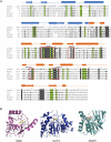
Figure 1.
Figure 1.
Dot1 is a conserved class I SAM-dependent methylase. (A) Sequence alignment of the conserved catalytic core region of Dot1 across multiple species that include yeast (NP_010728), worm (NM_058569), fly (AE003675), mouse (BB678539), and human (NP_115871). Secondary structure elements are shown above the alignment. The dashed line indicates disordered regions. Invariant amino acids are shown in white letters against a gray background, while conserved residues are in bold. Amino acids conserved across all five species are in bold against a green background. Every 10 amino acids is marked by a plus sign. The conserved methyltransferase fold motifs—I, post I, II, and III—are labeled and boxed in red. (B) The crystal structure of the substrate-binding pocket of human DOT1L (Protein Data Bank [PDB] code 1NW3) demonstrates that it is more similar to that of rat PRMT1 (PDB code 1ORI), a class I arginine methyltransferase, than to the KMT hNSD1 (PDB code 3OOI).
Figure 2.
Figure 2.
Ubiquitination of yH2B-K123/hH2B-K120 regulates Dot1-mediated H3K79 methylation. (A) The crystal structure of the human nucleosome (PDB code 3AFA) is shown on the left. Histone H2A is shown in light green, H4 is shown in sand, H2B is shown in hot pink, and H3 is shown in blue. A close-up view of the nucleosome surface is on the right, with the location of H2B-K120 shown as yellow spheres and H3K79 shown as green spheres. Note that H2B-K120 and H3K79 are located in proximity on the same solvent-exposed surface of the nucleosome. (B) Proposed models for trans-histone regulation of Dot1 enzymatic activity by H2B-K123ub. Histone H2B-K123 ubiquitination is mediated by the ubiquitin-conjugating enzyme Rad6 and its E3 ubiquitin ligase, Bre1. Components of the Paf1 complex are required for efficient H2B ubiquitination and di- and trimethylation of H3K4 and H3K79. (Bottom left) The first model suggests that Dot1 can bind directly to H2B-K123ub for intranucleosomal H3K79 methylation. (Top right) The second model suggests that other factors, such as the Set1-containing COMPASS, are required to bridge the interaction between Dot1 and the nucleosome. Rtf1 of the Paf1 complex recruits the COMPASS complex to methylate H3K4. The Cps35 subunit of the COMPASS interacts directly with H2B-K123 and Dot1, thus bringing Dot1 to the nucleosome for H3K79 methylation. (Bottom right) In the third model, ubiquitination of H2B causes a conformational change to the nucleosome, making H3K79 more accessible to DOT1.
Figure 3.
Figure 3.
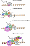
Mechanism for the involvement of DOT1L-mediated H3K79 methylation in transcriptional elongation. For simplicity of the model, only a fraction of the proteins involved in RNA Pol II transcription is shown. In step I, RNA Pol II is phosphorylated on Ser5 of its CTD and has initiated transcription. Negative elongation factor (NELF) and 5,6-dichloro-1b-D-ribofuranosylbenzimidazole sensitivity-inducing factor (DSIF) are bound to RNA Pol II for promoter-proximal pausing. In step II, promoter clearance is achieved by the recruitment of P-TEFb, which phosphorylates Ser2 of the RNA Pol II CTD, NELF, and DSIF. Phosphorylated NELF dissociates to release Pol II. The PAF1 complex, along with RAD6 and BRE1, is recruited to ubiquitinate H2B-K120. In step III, which can occur simultaneously with step II, H3K4 di- and trimethylation is achieved by the recruitment of MLL, the human homolog of Set1, by the PAF1 complex and association with H2B-K120ub. During transcription elongation shown in step IV, DOT1L is recruited to elongating RNA Pol II through its association with a network of proteins that include, but are not limited to, the elongation factors ELL, AF4, AF10, AF9, and ENL. DOT1L methylates H3K79 to maintain active transcription and/or serve as a transcription memory.
Figure 4.
Figure 4.
Role of Dot1 in DNA damage checkpoint response and repair. The yeast proteins are shown, and the steps in which Dot1 has been implicated are marked by red stars. (A) In yeast, the PI-3 kinases Mec1/Ddc2 (human ATR/ATRIP) sense exposed ssDNA formed during repair of UV-, oxidative-, and alkylating-damaged DNA as well as resected DSBs. The sensor PI-3 kinase Tel (human ATM) recognizes blunt-end DSBs. After recognition and binding of sensor proteins to the site of DNA damage, mediator proteins such as Rad9, Dpb11, and Mrc1 are recruited and activated by phosphorylation. Dot1-mediated H3K79 methylation is required for the recruitment and activation of Rad9 and phosphorylation of the downstream transducer protein Rad53. Dot1 is also required for proper checkpoint function at both G1 and G2. (B) Several different DNA repair mechanisms can be used by the cell in response to different types of DNA damage. Damage by UV radiation can be repaired by NER, RR, and PRR. Loss of H3K79 methylation impairs all three pathways, resulting in hypersensitivity to UV damage. Damage by alkylating agents can be repaired by the BER pathway. In rare instances where the damage cannot be repaired, the error-prone TLS pathway can be activated to bypass the DNA lesion. It was demonstrated that Dot1 maintains DNA integrity by inhibiting TLS. Finally, IR, which generates DSBs, can be repaired by NHEJ. Alternatively, the DSBs can undergo 5′-to-3′ resection and be repaired by HR. Methylation of H3K79 and the subsequent recruitment of Rad9 are required for regulating resection. Additionally, H3K79 methylation is required for the recruitment of cohesin, which is important for maintaining chromosome structure and for efficient SCR to repair DSBs.
Figure 5.
Figure 5.
DOT1L contributes to cardiac function by activating dystrophin expression. DOT1L-mediated H3K79 methylation is required for dystrophin expression. Dystrophin forms a DGC that plays a pivotal role in lateral force transduction and relieving mechanical stress for optimal cardiac performance. Loss of H3K79 methylation results in silencing of the dystrophin locus, loss of expression of the DGC complex, and sarcolemma damage. The absence of a functional DGC is manifested by systolic dysfunction and DCM.
Figure 6.
Figure 6.
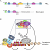
MLL fusion proteins recruit DOT1L for transcriptional activation and leukemogenesis. (A, top) MLL is a SET domain-containing H3K4 methyltransferase with multiple functional domains. (AT-hook and CxxC) DNA binding; (SNL) signal nuclear localization; (PHD) protein interactions; (TAD) transactivation domain; (SET) methyltransferase domain. Frequent DSBs occur within the MLL locus at a breakpoint cluster region (BCR). (Bottom) The N terminus of MLL undergoes chromosomal rearrangements and fuses in-frame to >50 different genes, resulting in the expression of leukemogenic MLL fusion proteins. (B) Constitutive activation of a leukemic transcriptional program, including Hoxa and Meis1 genes, is achieved through mistargeting of DOT1L and its associated proteins through the interaction of DOT1L with MLL fusion partners. DOT1L-mediated H3K79 methylation and the subsequent activation of the target genes result in leukemic transformation.

References

    1. Altaf M, Utley RT, Lacoste N, Tan S, Briggs SD, Cote J 2007. Interplay of chromatin modifiers on a short basic patch of histone H4 tail defines the boundary of telomeric heterochromatin. Mol Cell 28: 1002–1014 - PMC - PubMed
    1. Ayton PM, Cleary ML 2001. Molecular mechanisms of leukemogenesis mediated by MLL fusion proteins. Oncogene 20: 5695–5707 - PubMed
    1. Barry ER, Krueger W, Jakuba CM, Veilleux E, Ambrosi DJ, Nelson CE, Rasmussen TP 2009. ES cell cycle progression and differentiation require the action of the histone methyltransferase Dot1L. Stem Cells 27: 1538–1547 - PMC - PubMed
    1. Barski A, Cuddapah S, Cui K, Roh TY, Schones DE, Wang Z, Wei G, Chepelev I, Zhao K 2007. High-resolution profiling of histone methylations in the human genome. Cell 129: 823–837 - PubMed
    1. Bitoun E, Oliver PL, Davies KE 2007. The mixed-lineage leukemia fusion partner AF4 stimulates RNA polymerase II transcriptional elongation and mediates coordinated chromatin remodeling. Hum Mol Genet 16: 92–106 - PubMed

Publication types

LinkOut - more resources