. 2011 Jul 1;25(13):1426-38.
doi: 10.1101/gad.2024411.

Fine-tuning p53 activity through C-terminal modification significantly contributes to HSC homeostasis and mouse radiosensitivity

Affiliations

Fine-tuning p53 activity through C-terminal modification significantly contributes to HSC homeostasis and mouse radiosensitivity

Yunyuan V Wang et al. Genes Dev. .

Abstract

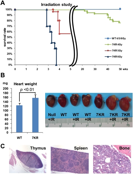
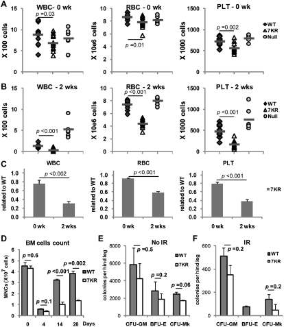
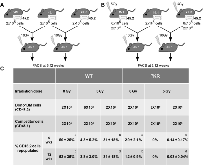
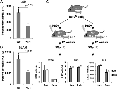
Cell cycle regulation in hematopoietic stem cells (HSCs) is tightly controlled during homeostasis and in response to extrinsic stress. p53, a well-known tumor suppressor and transducer of diverse stress signals, has been implicated in maintaining HSC quiescence and self-renewal. However, the mechanisms that control its activity in HSCs, and how p53 activity contributes to HSC cell cycle control, are poorly understood. Here, we use a genetically engineered mouse to show that p53 C-terminal modification is critical for controlling HSC abundance during homeostasis and HSC and progenitor proliferation after irradiation. Preventing p53 C-terminal modification renders mice exquisitely radiosensitive due to defects in HSC/progenitor proliferation, a critical determinant for restoring hematopoiesis after irradiation. We show that fine-tuning the expression levels of the cyclin-dependent kinase inhibitor p21, a p53 target gene, contributes significantly to p53-mediated effects on the hematopoietic system. These results have implications for understanding cell competition in response to stresses involved in stem cell transplantation, recovery from adverse hematologic effects of DNA-damaging cancer therapies, and development of radioprotection strategies.


Figures

Figure 1.
Figure 1.
Preventing C-terminal modification of p53 engenders exquisite radiosensitivity, particularly in the hematopoietic system. (A) Kaplan-Meier radiation survival curves in WT mice and p537KR (7KR) mice after 4, 5, and 6 Gy of whole-body irradiation showing dose-dependent mortality in p537KR mice (4 Gy: n = 58, P < 0.001; 5 Gy: n = 25, P < 0.001; 6 Gy: n = 10, P < 0.001). (B) Increased cardiac weights and enlarged hearts in irradiated p537KR mice 4 wk after 5 Gy of irradiation. Error bars represent the SEM from four animals. The representative hearts from nonirradiated and irradiated mice are shown in the picture. (C) H&E staining of thymus, spleen, and BM from a p537KR mouse that died after exposure to 5 Gy of whole-body irradiation.
Figure 2.
Figure 2.
Irradiation causes long-term defects in p537KR hematopoietic progenitor cells. (A) White blood cells (WBC) red blood cells (RBC), and and platelets (PLT) in WT, p537KR, and p53-null mice prior to irradiation. WT and 7KR: n = 13; null: n = 6. (B) Blood cell counts at 2 wk after 5 Gy of whole-body irradiation. (C) The fold changes of blood cell counts in p537KR mice related to WT mice 2 wk post-irradiation. (D) Mononucleated cell (MNCs) count in BM at baseline and 4, 14, and 28 d post-irradiation revealed insufficient recovery of BM cells in irradiated p537KR mice. (E) BM cells were isolated from nonirradiated mice and seeded for granulocytic/macrophage (CFU-GM), erythroid (BFU-E), and megakaryocytic (CFU-MK) progenitor cell colony-forming assay. (F) BM cells were immediately isolated from animals that were exposed to irradiation for progenitor colony-forming assay. Note: No colonies were found when 5 × 10⁶ p537KR BM cells were seeded for BFU-E assay. The error bars in D, E, and F represent SEM from three independent experiments.
Figure 3.
Figure 3.
p537KR HSCs exhibit sensitivity to transplantation and to irradiation. BM from nonirradiated (A) and 4-d post-irradiated (B) WT or p537KR donor mice expressing the CD45.2 leukocyte cell surface marker were transplanted into lethally irradiated recipient mice in fixed ratios with WT competitor marrow cells expressing CD45.1. (C) p537KR HSCs did not compete as well as WT for long-term repopulation under both nonirradiated and irradiated conditions. At 6 wk and 12 wk post-transplantation, peripheral blood was withdrawn from the recipients and analyzed for the percentage of CD45.2 cells in total leukocytes. Results show mean ± SD from at least five animals. a, b: P < 0.05. c, d: P < 0.001.
Figure 4.
Figure 4.
The hematopoietic defects in p537KR mice reside in HSCs/progenitor cells and not in the stem cell niche. (A) BM cells were isolated from nonirradiated mice and stained with antibodies against hematopoietic lineage (Lin) markers and cell surface marks Sca1 and c-Kit and analyzed by flow cytometry. LinSca1+c-Kit+ subpopulation were gated as HSC-enriched LSK cells. Cell plots are shown in Supplemental Figure S4. (B) Antibody cocktail lineage markers CD41, CD48, and CD150 were used for the analysis. The LinCD41CD48CD150+ subpopulation was gated as HSC-enriched SLAM cells. Cell plots are shown in Supplemental Figure S4. (C) Introducing WT HSCs into p537KR mice by BM transplantation overcame the radiosensitivity of the animals. BM cells expressing CD45.1 were transplanted into lethally irradiated WT or p537KR mice. Twelve weeks later, the chimeric mice were exposed to 5 Gy of irradiation followed by CBC analysis. (A–C) Error bars represent the SEM from three animals.
Figure 5.
Figure 5.
Mdm2 dependence of radiosensitivity in p537KR mice. (A) Kaplan-Meier radiation survival curves. WT;Mdm2+/− (n = 9) and p537KR;Mdm2+/− (n = 6) mice under the same genetic background were exposed to 5 Gy whole-body irradiation. Losing one allele of Mdm2 enhanced the radiosensitivity of p537KR mice. P < 0.001. (B) CBC analysis of p537KR;Mdm2+/+ and p537KR;Mdm2+/− mice before irradiation (n = 9) and 2 wk after 5 Gy whole-body irradiation (n = 5) showed severe pancytopenia in p537KR;Mdm2+/− mice. (C) Histopathological evaluation revealed that p537KR;Mdm2+/− mice that died from irradiation exhibited severe atrophy in the spleen and BM. (D) Western analysis of p53 and p21 abundance in spleens isolated from WT, Mdm2+/− (M2+/−), p537KR (7KR), and p537KR;Mdm2+/− (7KR;M2+/−) mice prior to or 3 h after 5 Gy of whole-body irradiation. Actin blot provides a loading control. Intensities of p53 and p21 signals were normalized to Actin signals and are presented as relative fold differences. (E) p537KR;Mdm2+/− mice exhibited a slightly lower frequency of HSC-enriched SLAM cells when compared with p537KR;Mdm2+/+ mice. Error bars represent the SEM from three animals.
Figure 6.
Figure 6.
Differential gene expression and cell cycle kinetics in p537KR mice. (A) The p53-inducible proapoptotic gene puma was expressed at higher levels in thymus than in BM after irradiation. The level of puma expression was similar between WT and p537KR mice. (B) The p53-inducible cell cycle arrest gene p21 was highly expressed in BM after irradiation, and showed even greater expression in p537KR animals. (A,B) Error bars represent the SEM from three animals. (C) Expression of 43 p53 target genes from thymus (T) and BM (B) prior to irradiation or 3 h post-irradiation (IR) was detected using microfluidic qPCR analysis. Relative expressions of each gene from all samples were scaled from 0 to 1, followed by hierarchical clustering analysis. (Cluster I) Repressive or less responsive genes. (Cluster II) Highly IR-induced genes in thymus. (Cluster III) Genes highly affected by 7KR mutations in BM. (Cluster IV) Highly IR-induced genes in BM. (Cluster V) Highly expressed genes in BM. The analysis suggests that several genes (highlighted in pink) are differentially affected by the 7KR mutations. (D) Thymi, spleens, and BM were isolated from mice at 0, 3, and 6 h after 5 Gy of whole-body irradiation. Apoptosis was detected by TUNEL staining (green). (E) Nonirradiated mice or mice exposed to 5 Gy of irradiation were injected with 2 mg of BrdU followed by 2-d administration of BrdU water (1 mg/mL). BMs were then isolated and stained with antibodies against lineage (Lin) markers; cell surface markers Sca1, c-kit, and CD34; and BrdU, followed by flow analysis. BrdU incorporated cell in Lin, LSK (LinSca1+c-kit+) and LSKCD34 subcell populations were analyzed. Error bars represent the SD from three animals.
Figure 7.
Figure 7.
p21 gene dosage affects radiosensitivity of p537KR mice. (A) Kaplan-Meier radiation survival curves. Littermate p537KR;p21+/+ (n = 7) and p537KR;p21+/− (n = 10) mice were exposed to 6 Gy of whole-body irradiation. Losing one allele of p21 partially rescued the sensitivity of p537KR mice to 6 Gy irradiation. P = 0.06. (B) Analysis of gene expression in thymus and BM showed that p21 expression was reduced by half in p537KR;p21+/− mice (p21+/−), while puma expression was not affected. Error bars represent the SEM from three animals. (C) Western analysis showed reduced p21 levels in irradiated p537KR;p21+/− mice, while p53 levels were unchanged. (D) p537KR;p21+/− mice exhibited slight lower frequency of HSC-enriched SLAM cells when compared with p537KR;p21+/+ mice. Error bars represent the SEM from three animals.

References

    1. Appella E, Anderson CW 2001. Post-translational modifications and activation of p53 by genotoxic stresses. Eur J Biochem 268: 2764–2772 - PubMed
    1. Aranda-Anzaldo A, Dent MAR 2007. Reassessing the role of p53 in cancer and ageing from an evolutionary perspective. Mech Ageing Dev 128: 293–302 - PubMed
    1. Bondar T, Medzhitov R 2010. p53-mediated hematopoietic stem and progenitor cell competition. Cell Stem Cell 6: 309–322 - PMC - PubMed
    1. Chen J, Felicia ME, Keyvan K, Stephanie OO, Marie JD, Michael AE, Neal SY 2008. Enrichment of hematopoietic stem cells with SLAM and LSK markers for the detection of hematopoietic stem cell function in normal and Trp53 null mice. Exp Hematol 36: 1236–1243 - PMC - PubMed
    1. Cheng T, Rodrigues N, Shen H, Yang Y-g, Dombkowski D, Sykes M, Scadden DT 2000. Hematopoietic stem cell quiescence maintained by p21cip1/waf1. Science 287: 1804–1808 - PubMed

Publication types

MeSH terms

Substances