Lipopolysaccharide-induced injury is more pronounced in fetal transgenic ErbB4-deleted lungs
- PMID: 21724861
- PMCID: PMC3191748
- DOI: 10.1152/ajplung.00131.2010
Lipopolysaccharide-induced injury is more pronounced in fetal transgenic ErbB4-deleted lungs
Abstract
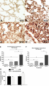
Pulmonary ErbB4 deletion leads to a delay in fetal lung development, alveolar simplification, and lung function disturbances in adult mice. We generated a model of intrauterine infection in ErbB4 transgenic mice to study the additive effects of antenatal LPS administration and ErbB4 deletion during fetal lung development. Pregnant mice were treated intra-amniotically with an LPS dose of 4 μg at E17 of gestation. Lungs were analyzed 24 h later. A significant influx of inflammatory cells was seen in all LPS-treated lungs. In heterozygote control lungs, LPS treatment resulted in a delay of lung morphogenesis characterized by a significant increase in the fraction of mesenchyme, a decrease in gas exchange area, and disorganization of elastic fibers. Surfactant protein (Sftp)b and Sftpc were upregulated, but mRNA of Sftpb and Sftpc was downregulated compared with non-LPS-treated controls. The mRNA of Sftpa1 and Sftpd was upregulated. In ErbB4-deleted lungs, the LPS effects were more pronounced, resulting in a further delay in morphological development, a more pronounced inflammation in the parenchyma, and a significant higher increase in all Sftp. The effect on Sftpb and Sftpc mRNA was somewhat different, resulting in a significant increase. These results imply a major role of ErbB4 in LPS-induced signaling in structural and functional lung development.
Figures


Similar articles
-
ErbB4 is an upstream regulator of TTF-1 fetal mouse lung type II cell development in vitro.Biochim Biophys Acta. 2013 Dec;1833(12):2690-2702. doi: 10.1016/j.bbamcr.2013.06.030. Epub 2013 Jul 8. Biochim Biophys Acta. 2013. PMID: 23845988 Free PMC article.
-
Bone marrow stem cells accelerate lung maturation and prevent the LPS-induced delay of morphological and functional fetal lung development in the presence of ErbB4.Cell Tissue Res. 2020 Jun;380(3):547-564. doi: 10.1007/s00441-019-03145-0. Epub 2020 Feb 13. Cell Tissue Res. 2020. PMID: 32055958
-
Neuregulin receptor ErbB4 functions as a transcriptional cofactor for the expression of surfactant protein B in the fetal lung.Am J Respir Cell Mol Biol. 2011 Oct;45(4):761-7. doi: 10.1165/rcmb.2010-0179OC. Epub 2011 Feb 11. Am J Respir Cell Mol Biol. 2011. PMID: 21317380 Free PMC article.
-
Presenilin-1 processing of ErbB4 in fetal type II cells is necessary for control of fetal lung maturation.Biochim Biophys Acta. 2011 Mar;1813(3):480-91. doi: 10.1016/j.bbamcr.2010.12.017. Epub 2010 Dec 29. Biochim Biophys Acta. 2011. PMID: 21195117 Free PMC article.
-
Alveolar-Capillary Membrane-Related Pulmonary Cells as a Target in Endotoxin-Induced Acute Lung Injury.Int J Mol Sci. 2019 Feb 15;20(4):831. doi: 10.3390/ijms20040831. Int J Mol Sci. 2019. PMID: 30769918 Free PMC article. Review.
Cited by
-
Can We Understand the Pathobiology of Bronchopulmonary Dysplasia?J Pediatr. 2017 Nov;190:27-37. doi: 10.1016/j.jpeds.2017.08.041. J Pediatr. 2017. PMID: 29144252 Free PMC article. Review. No abstract available.
-
Loss of lung WWOX expression causes neutrophilic inflammation.Am J Physiol Lung Cell Mol Physiol. 2017 Jun 1;312(6):L903-L911. doi: 10.1152/ajplung.00034.2017. Epub 2017 Mar 10. Am J Physiol Lung Cell Mol Physiol. 2017. PMID: 28283473 Free PMC article.
-
Surfactant Protein D in Respiratory and Non-Respiratory Diseases.Front Med (Lausanne). 2018 Feb 8;5:18. doi: 10.3389/fmed.2018.00018. eCollection 2018. Front Med (Lausanne). 2018. PMID: 29473039 Free PMC article. Review.
-
Cigarette Smoke and Nicotine-Containing Electronic-Cigarette Vapor Downregulate Lung WWOX Expression, Which Is Associated with Increased Severity of Murine Acute Respiratory Distress Syndrome.Am J Respir Cell Mol Biol. 2021 Jan;64(1):89-99. doi: 10.1165/rcmb.2020-0145OC. Am J Respir Cell Mol Biol. 2021. PMID: 33058734 Free PMC article.
-
Intra-amniotic LPS and antenatal betamethasone: inflammation and maturation in preterm lamb lungs.Am J Physiol Lung Cell Mol Physiol. 2012 Feb 15;302(4):L380-9. doi: 10.1152/ajplung.00338.2011. Epub 2011 Dec 9. Am J Physiol Lung Cell Mol Physiol. 2012. PMID: 22160306 Free PMC article.
References
-
- Anderson L, Seilhamer J. A comparison of selected mRNA and protein abundances in human liver. Electrophoresis 18: 533–537, 1997 - PubMed
-
- Arias-Diaz J, Garcia-Verdugo I, Casals C, Sanchez-Rico N, Vara E, Balibrea JL. Effect of surfactant protein A (SP-A) on the production of cytokines by human pulmonary macrophages. Shock 14: 300–306, 2000 - PubMed
-
- Bachurski CJ, Ross GF, Ikegami M, Kramer BW, Jobe AH. Intra-amniotic endotoxin increases pulmonary surfactant proteins and induces SP-B processing in fetal sheep. Am J Physiol Lung Cell Mol Physiol 280: L279–L285, 2001 - PubMed
-
- Bell MJ, Hallenbeck JM, Gallo V. Determining the fetal inflammatory response in an experimental model of intrauterine inflammation in rats. Pediatr Res 56: 541–546, 2004 - PubMed
-
- Beloosesky R, Gayle DA, Amidi F, Nunez SE, Babu J, Desai M, Ross MG. N-acetyl-cysteine suppresses amniotic fluid and placenta inflammatory cytokine responses to lipopolysaccharide in rats. Am J Obstet Gynecol 194: 268–273, 2006 - PubMed
Publication types
MeSH terms
Substances
Grants and funding
LinkOut - more resources
Full Text Sources
Molecular Biology Databases
Miscellaneous

