

Diastolic dysfunction is associated with cardiac fibrosis in the senescence-accelerated mouse
- PMID: 21724869
- PMCID: PMC3191096
- DOI: 10.1152/ajpheart.00407.2010
Diastolic dysfunction is associated with cardiac fibrosis in the senescence-accelerated mouse
Abstract
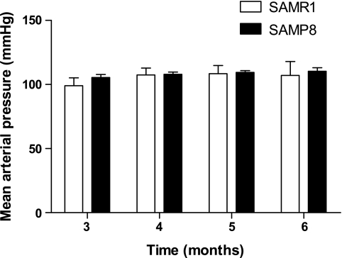
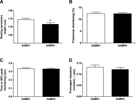
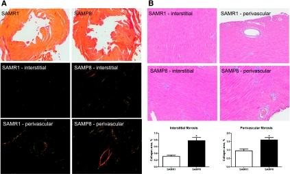
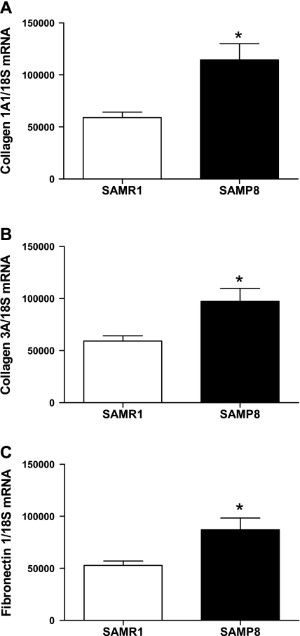
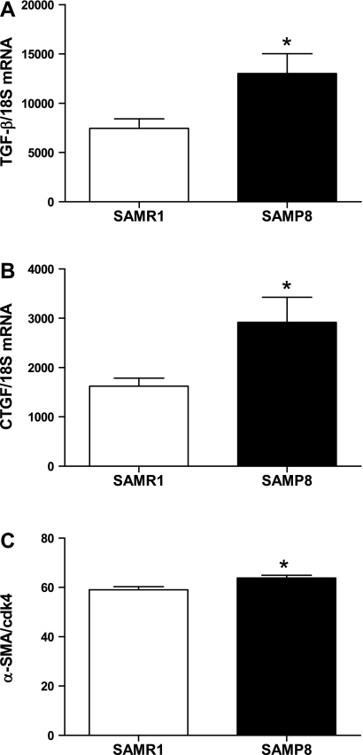
Diastolic heart failure is a major cause of mortality in the elderly population. It is often preceded by diastolic dysfunction, which is characterized by impaired active relaxation and increased stiffness. We tested the hypothesis that senescence-prone (SAMP8) mice would develop diastolic dysfunction compared with senescence-resistant controls (SAMR1). Pulsed-wave Doppler imaging of the ratio of blood flow velocity through the mitral valve during early (E) vs. late (A) diastole was reduced from 1.3 ± 0.03 in SAMR1 mice to 1.2 ± 0.03 in SAMP8 mice (P < 0.05). Tissue Doppler imaging of the early (E') and late (A') diastolic mitral annulus velocities found E' reduced from 25.7 ± 0.9 mm/s in SAMR1 to 21.1 ± 0.8 mm/s in SAMP8 mice and E'/A' similarly reduced from 1.1 ± 0.02 to 0.8 ± 0.03 in SAMR1 vs. SAMP8 mice, respectively (P < 0.05). Invasive hemodynamics revealed an increased slope of the end-diastolic pressure-volume relationship (0.5 ± 0.05 vs. 0.8 ± 0.14; P < 0.05), indicating increased left ventricular chamber stiffness. There were no differences in systolic function or mean arterial pressure; however, diastolic dysfunction was accompanied by increased fibrosis in the hearts of SAMP8 mice. In SAMR1 vs. SAMP8 mice, interstitial collagen area increased from 0.3 ± 0.04 to 0.8 ± 0.09% and perivascular collagen area increased from 1.0 ± 0.11 to 1.6 ± 0.14%. Transforming growth factor-β and connective tissue growth factor gene expression were increased in the hearts of SAMP8 mice (P < 0.05 for all data). In summary, SAMP8 mice show increased fibrosis and diastolic dysfunction similar to those seen in humans with aging and may represent a suitable model for future mechanistic studies.
Figures
References
-
- Aurigemma GP, Gaasch WH. Clinical practice. Diastolic heart failure. N Engl J Med 351: 1097–1105, 2004 - PubMed
-
- Basu R, Oudit GY, Wang X, Zhang L, Ussher JR, Lopaschuk GD, Kassiri Z. Type 1 diabetic cardiomyopathy in the Akita (Ins2WT/C96Y) mouse model is characterized by lipotoxicity and diastolic dysfunction with preserved systolic function. Am J Physiol Heart Circ Physiol 297: H2096–H2108, 2009 - PubMed
-
- Billet S, Bardin S, Verp S, Baudrie V, Michaud A, Conchon S, Muffat-Joly M, Escoubet B, Souil E, Hamard G, Bernstein KE, Gasc JM, Elghozi JL, Corvol P, Clauser E. Gain-of-function mutant of angiotensin II receptor, type 1A, causes hypertension and cardiovascular fibrosis in mice. J Clin Invest 117: 1914–1925, 2007 - PMC - PubMed
-
- Borlaug BA, Kass DA. Mechanisms of diastolic dysfunction in heart failure. Trends Cardiovasc Med 16: 273–279, 2006 - PubMed
Publication types
MeSH terms
Substances
Grants and funding
LinkOut - more resources
Full Text Sources
Medical