. 2011 Sep;77(17):5973-80.
doi: 10.1128/AEM.00253-11. Epub 2011 Jul 1.

Involvement of the pleiotropic drug resistance response, protein kinase C signaling, and altered zinc homeostasis in resistance of Saccharomyces cerevisiae to diclofenac

Affiliations

Involvement of the pleiotropic drug resistance response, protein kinase C signaling, and altered zinc homeostasis in resistance of Saccharomyces cerevisiae to diclofenac

Jolanda S van Leeuwen et al. Appl Environ Microbiol. 2011 Sep.

Abstract

Diclofenac is a widely used analgesic drug that can cause serious adverse drug reactions. We used Saccharomyces cerevisiae as a model eukaryote with which to elucidate the molecular mechanisms of diclofenac toxicity and resistance. Although most yeast cells died during the initial diclofenac treatment, some survived and started growing again. Microarray analysis of the adapted cells identified three major processes involved in diclofenac detoxification and tolerance. In particular, pleiotropic drug resistance (PDR) genes and genes under the control of Rlm1p, a transcription factor in the protein kinase C (PKC) pathway, were upregulated in diclofenac-adapted cells. We tested if these processes or pathways were directly involved in diclofenac toxicity or resistance. Of the pleiotropic drug resistance gene products, the multidrug transporter Pdr5p was crucially important for diclofenac tolerance. Furthermore, deletion of components of the cell wall stress-responsive PKC pathway increased diclofenac toxicity, whereas incubation of cells with the cell wall stressor calcofluor white before the addition of diclofenac decreased its toxicity. Also, diclofenac induced flocculation, which might trigger the cell wall alterations. Genes involved in ribosome biogenesis and rRNA processing were downregulated, as were zinc-responsive genes. Paradoxically, deletion of the zinc-responsive transcription factor Zap1p or addition of the zinc chelator 1,10-phenanthroline significantly increased diclofenac toxicity, establishing a regulatory role for zinc in diclofenac resistance. In conclusion, we have identified three new pathways involved in diclofenac tolerance in yeast, namely, Pdr5p as the main contributor to the PDR response, cell wall signaling via the PKC pathway, and zinc homeostasis, regulated by Zap1p.


Figures

Fig. 1.
Fig. 1.
Yeast cells can adapt to diclofenac. W303 cells were grown in the absence (squares) or presence (triangles) of diclofenac at 100 μM in minimal medium. After 24 and 48 h, the cultures were diluted in minimal medium containing no drug or 100 μM diclofenac. Growth is expressed as the OD600 ± SD.
Fig. 2.
Fig. 2.
Diclofenac-adapted cells have lower ROS levels than WT cells in the presence of diclofenac. W303 cells that either were not pretreated (WT) or were pretreated for 48 h with 100 μM diclofenac (adapted) were grown for 3 h with no drug (open bars) or 50 μM diclofenac (filled bars) in the presence of the ROS-sensitive fluorescent probe 2′,7′-dichlorodihydrofluorescein diacetate at 10 μM. Data are expressed as fluorescence units corrected for cell density (relative fluorescence units [RFU]) ± SD.
Fig. 3.
Fig. 3.
Adaptation to diclofenac is reversible. W303 strains either were grown in the absence of diclofenac (squares), pretreated for 72 h with 50 μM diclofenac and subsequently incubated with 150 μM diclofenac (circles), or first pretreated for 72 h with 50 μM diclofenac, then incubated for 48 h without diclofenac, and finally incubated with 150 μM diclofenac (triangles). Growth is expressed as the OD600 ± SD.
Fig. 4.
Fig. 4.
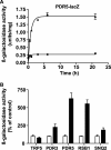
The pleiotropic drug response (PDR) is dramatically upregulated by diclofenac. (A) Time-dependent expression of PDR5-lacZ in a BY4741 strain incubated with no drug (squares) or with 30 μM diclofenac (triangles). Data are expressed as β-galactosidase activity units corrected for the protein concentration ± SD. (B) TRP5-, PDR3-, PDR5-, RSB1-, and SNQ2-lacZ expression in wild-type BY4741 cells incubated 3 h with no drug (open bars) or with 30 μM diclofenac (filled bars). LacZ expression is presented as a percentage of the β-galactosidase activity for untreated controls (set at100%) ± SD.
Fig. 5.
Fig. 5.
Pdr5p is important for diclofenac tolerance. BY4741 wild-type (open squares), Δpdr5 (open triangles), and PDR5-overexpressing (open circles) cells were grown in the presence of 100 μM diclofenac in minimal medium containing glucose. In the absence of diclofenac, wild-type (filled squares), Δpdr5 (not shown), and PDR5-overexpressing (not shown) cells grew comparably to each other. Data are expressed as the OD600 ± SD.
Fig. 6.
Fig. 6.
Diclofenac induces PKC pathway-mediated cell wall stress and flocculation. (A and B) Wild-type (filled symbols), Δslt2 (open symbols) (A), and Δpkc1 (open symbols) (B) W303 cells were incubated with no drug (squares) or with 100 μM diclofenac (triangles) in minimal medium containing 1 M sorbitol for osmostabilization. (C) Wild-type BY4741 cells were grown in the absence (squares) or presence (triangles) of 100 μM diclofenac in minimal medium containing no additives (filled symbols) or 100 μg/ml calcofluor white (CFW) (open symbols). Growth is expressed as the OD600 ± SD. (D) BY4741 WT and Δflo1 cells were grown in minimal medium in a 48-well plate. Photographs were taken 20 min after the addition of 50, 100, or 150 μM diclofenac or after addition of DMSO only (0 μM).
Fig. 7.
Fig. 7.
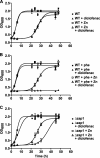
Diclofenac toxicity is increased under low-zinc conditions. Wild-type (A and B) or Δzap1 (C) BY4741 cells were incubated with no drug (squares) or with 100 μM diclofenac (triangles) in minimal medium. (A) The medium contained either no additive (filled symbols) or 50 μM ZnSO4 (open symbols). (B) The medium contained either 50 μM 1,10-phenanthroline (phe) alone (filled symbols) or both 50 μM ZnSO4 and 50 μM 1,10-phenanthroline (open symbols). (C) The medium contained either no additive (filled symbols) or 100 μM ZnSO4 (open symbols). Data are expressed as the OD600 ± SD.

References

    1. Abbott D. A., Suir E., van Maris A. J., Pronk J. T. 2008. Physiological and transcriptional responses to high concentrations of lactic acid in anaerobic chemostat cultures of Saccharomyces cerevisiae. Appl. Environ. Microbiol. 74:5759–5768 - PMC - PubMed
    1. Abou-Mohamed G., el-Kashef H. A., Salem H. A., Elmazar M. M. 1995. Effect of zinc on the anti-inflammatory and ulcerogenic activities of indometacin and diclofenac. Pharmacology 50:266–272 - PubMed
    1. Alem M. A., Douglas L. J. 2004. Effects of aspirin and other nonsteroidal anti-inflammatory drugs on biofilms and planktonic cells of Candida albicans. Antimicrob. Agents Chemother. 48:41–47 - PMC - PubMed
    1. Boorsma A., Foat B. C., Vis D., Klis F., Bussemaker H. J. 2005. T-profiler: scoring the activity of predefined groups of genes using gene expression data. Nucleic Acids Res. 33:W592–W595 - PMC - PubMed
    1. Carvajal E., van den Hazel H. B., Cybularz-Kolaczkowska A., Balzi E., Goffeau A. 1997. Molecular and phenotypic characterization of yeast PDR1 mutants that show hyperactive transcription of various ABC multidrug transporter genes. Mol. Gen. Genet. 256:406–415 - PubMed

MeSH terms