

doi: 10.1128/JB.05028-11.
Epub 2011 Jul 1.
The sulfolobicin genes of Sulfolobus acidocaldarius encode novel antimicrobial proteins
Affiliations
- PMID: 21725003
- PMCID: PMC3165506
- DOI: 10.1128/JB.05028-11
Item in Clipboard
The sulfolobicin genes of Sulfolobus acidocaldarius encode novel antimicrobial proteins
J Bacteriol.
2011 Sep.
Abstract
Crenarchaea, such as Sulfolobus acidocaldarius and Sulfolobus tokodaii, produce antimicrobial proteins called sulfolobicins. These antimicrobial proteins inhibit the growth of closely related species. Here we report the identification of the sulfolobicin-encoding genes in S. acidocaldarius. The active sulfolobicin comprises two proteins that are equipped with a classical signal sequence. These proteins are secreted by the cells and found to be membrane vesicle associated. Gene inactivation studies demonstrate that both proteins are required for the bacteriostatic antimicrobial activity. Sulfolobicins constitute a novel class of antimicrobial proteins without detectable homology to any other protein.
Figures
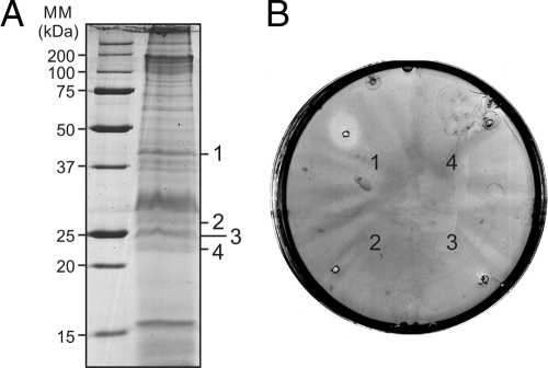
Antimicrobial activity of proteins isolated from membrane vesicles derived from Sulfolobus tokodaiicells. (A) S. tokodaiivesicles were loaded on an SDS-polyacrylamide gel, and proteins were separated. Protein bands were excised from the gel. The protein bands were numbered 1 to 4 and are shown to the right of the gel. The positions of molecular mass markers (MM) (in kilodaltons) are shown to the left of the gel. (B) The protein bands excised from the gel were loaded on plates containing a lawn of Sulfolobus solfataricusstrain P1 reporter cells. The active slice (band 1) was analyzed by LC-MS.
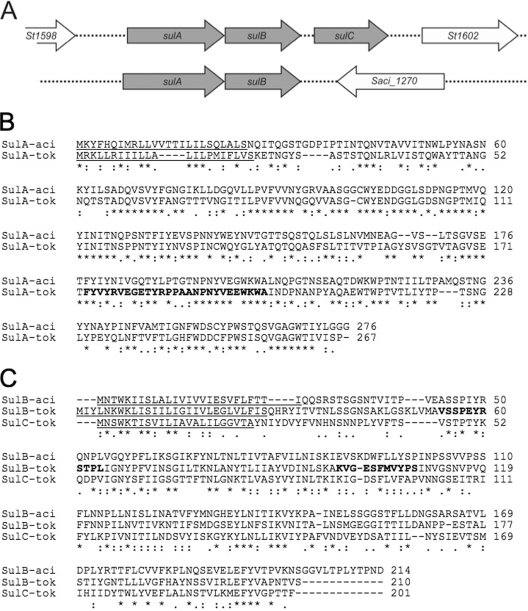
Genomic organization and amino acid sequences of the sulfolobicin genes. (A) Genomic organization of the sulfolobicin genes of Sulfolobus tokodaii(top line) and Sulfolobus acidocaldarius(bottom line). The S. tokodaii sulCgene appears to be a duplication of sulBbut is absent from the S. acidocaldariusgenome. (B and C) ClustalW2 alignment of SulA (B) and SulBC (C) proteins from S. acidocaldariusDSM 639 (-aci) and S. tokodaiistrain 7 (-tok). Predicted signal peptides are underlined, and peptides identified by LC-MS are depicted in bold type. Gaps introduced to maximize alignment are indicated by dashes. Amino acids that are identical in the different sequences are indicated by an asterisk below the sequence alignment. For S. tokodaii, the sulAgene is also called St1599, the sulBgene is also called St1600, and the sulCgene is also called St1601. For S. acidocaldarius, the sulAgene is also called Saci_1271, and the sulBgene is also called Saci_1272.
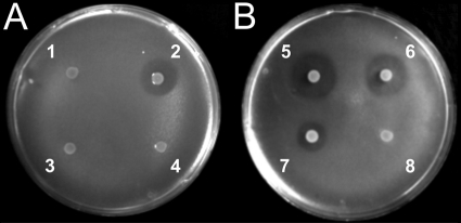
Overlay assay with colonies of S. acidocaldariusMW001 grown on a lawn of S. solfataricusstrain P2. The S. acidocaldariusMW001 strains used are indicated by the following numbers on the plates: 1, ΔsulABmutant; 2, wild type; 3, ΔsulBmutant; 4, ΔsulAmutant; 5 and 6, ΔsulABmutant complemented with sulAB[MW001 (sulAB)]; 7, wild type; and 8, ΔsulABmutant.
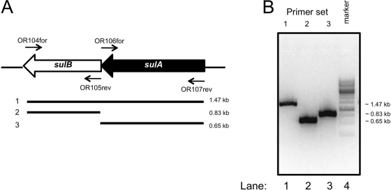
The sulAand sulBgenes are coexpressed from a single transcript. (A) Positions of the primers used to detect cDNA fragments and the expected PCR fragments. (B) cDNA was synthesized from RNA isolated from S. acidocaldariusMW001 cells grown to late exponential phase (OD600of ∼0.7). The indicated primer sets were used to detect the presence of PCR products. When reverse transcriptase was left out of the cDNA synthesis reaction, no PCR products were detected.
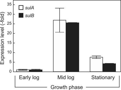
Expression levels of the sulABgenes from S. acidocaldariusMW001 during the indicated growth stages. Early log, mid log, and stationary growth phase correspond to OD600s of 0.1, 0.5, and 1.5, respectively. Expression was determined by real-time quantitative PCR and normalized relative to the expression of sulAand sulBin the early log phase with the secYgene as a reference.
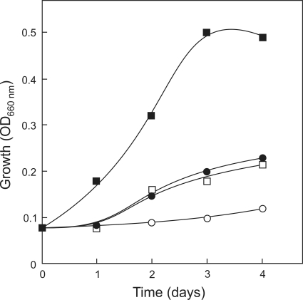
Effect of sulfolobicin produced by S. acidocaldariusMW001 on the growth of S. solfataricusP2 in liquid culture. Supernatant from medium in which wild-type S. acidocaldarius(0 to 50 ml) and ΔsulABcells (0 to 50 ml) was grown (spent medium) was mixed with a growing culture of S. solfataricusP2 (25 ml). Next, growth was monitored over time by measuring the OD660. Symbols: ▪, 50 ml of ΔsulABmutant supernatant (control); ○, 50 ml of wild-type strain MW001 supernatant; □, 25 ml (each) of supernatant from the ΔsulABmutant and MW001 strain; •, 15 ml of supernatant from strain MW001 and 35 ml of supernatant from ΔsulABmutant.
References
-
- Brock T. D., Brock K. M., Belly R. T., Weiss R. L. 1972. Sulfolobus: a new genus of sulfur-oxidizing bacteria living at low pH and high temperature. Arch. Mikrobiol. 84:54–68 - PubMed
-
- Contursi P., et al. 2006. Characterization of the Sulfolobus host-SSV2 virus interaction. Extremophiles 10:615–627 - PubMed
-
- DeLong E. F., Pace N. R. 2001. Environmental diversity of bacteria and archaea. Syst. Biol. 50:470–478 - PubMed
Publication types
MeSH terms
Substances
LinkOut - more resources
Full Text Sources
Other Literature Sources
Molecular Biology Databases
