

Verification of computed tomographic estimates of cochlear implant array position: a micro-CT and histologic analysis
- PMID: 21725264
- PMCID: PMC3173021
- DOI: 10.1097/MAO.0b013e3182255915
Verification of computed tomographic estimates of cochlear implant array position: a micro-CT and histologic analysis
Abstract
Objective: To determine the efficacy of clinical computed tomographic (CT) imaging to verify postoperative electrode array placement in cochlear implant (CI) patients.
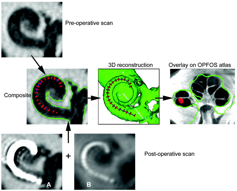
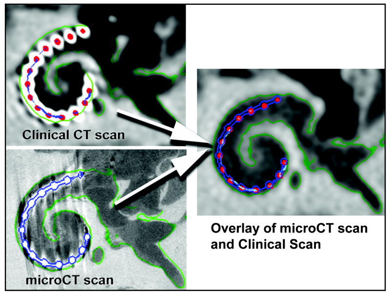
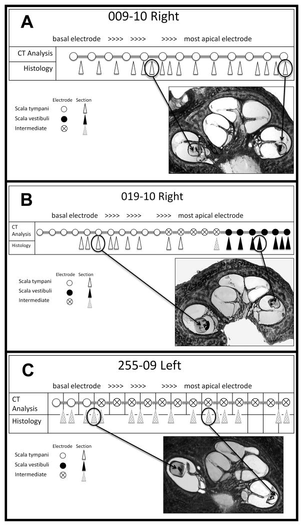
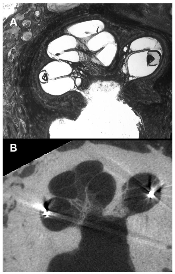
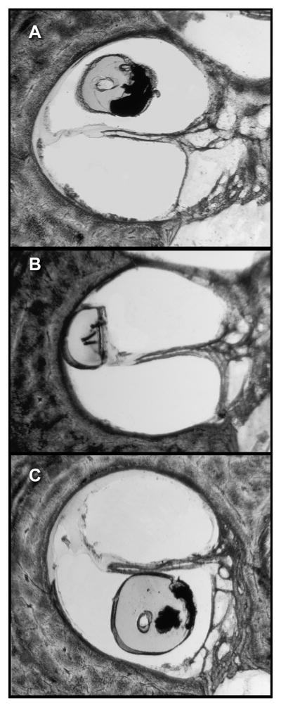
Study design: Nine fresh cadaver heads underwent clinical CT scanning, followed by bilateral CI insertion and postoperative clinical CT scanning. Temporal bones were removed, trimmed, and scanned using micro-CT. Specimens were then dehydrated, embedded in either methyl methacrylate or LR White resin, and sectioned with a diamond wafering saw. Histology sections were examined by 3 blinded observers to determine the position of individual electrodes relative to soft tissue structures within the cochlea. Electrodes were judged to be within the scala tympani, scala vestibuli, or in an intermediate position between scalae.
Results: The position of the array could be estimated accurately from clinical CT scans in all specimens using micro-CT and histology as a criterion standard. Verification using micro-CT yielded 97% agreement, and histologic analysis revealed 95% agreement with clinical CT results.
Conclusion: A composite, 3-dimensional image derived from a patient's preoperative and postoperative CT images using a clinical scanner accurately estimates the position of the electrode array as determined by micro-CT imaging and histologic analyses. Information obtained using the CT method provides valuable insight into numerous variables of interest to patient performance such as surgical technique, array design, and processor programming and troubleshooting.
Figures
References
-
- Balkany TJ, Connell SS, Hodges AV, et al. Conservation of residual acoustic hearing after cochlear implantation. Otol Neurotol. 2006;27:1083–8. - PubMed
-
- Fraysse B, Macias AR, Sterkers O, et al. Residual hearing conservation and electroacoustic stimulation with the nucleus 24 contour advance cochlear implant. Otol Neurotol. 2006;27:624–33. - PubMed
-
- Gantz BJ, Turner C, Gfeller KE, et al. Preservation of hearing in cochlear implant surgery: advantages of combined electrical and acoustical speech processing. Laryngoscope. 2005;115:796–802. - PubMed
-
- James CJ, Fraysse B, Deguine O, et al. Combined electroacoustic stimulation in conventional candidates for cochlear implantation. Audiol Neurootol. 2006;11(Suppl 1):57–62. - PubMed
Publication types
MeSH terms
Grants and funding
LinkOut - more resources
Full Text Sources
