. 2011 Jul 3;7(8):531-7.
doi: 10.1038/nchembio.598.

Small-molecule displacement of a cryptic degron causes conditional protein degradation

Affiliations

Small-molecule displacement of a cryptic degron causes conditional protein degradation

Kimberly M Bonger et al. Nat Chem Biol. .

Abstract

The ability to rapidly regulate the functions of specific proteins in living cells is a valuable tool for biological research. Here we describe a new technique by which the degradation of a specific protein is induced by a small molecule. A protein of interest is fused to a ligand-induced degradation (LID) domain, resulting in the expression of a stable and functional fusion protein. The LID domain is comprised of the FK506- and rapamycin-binding protein (FKBP) and a 19-amino-acid degron fused to the C terminus of FKBP. In the absence of the small molecule Shield-1, the degron is bound to the FKBP fusion protein and the protein is stable. When present, Shield-1 binds tightly to FKBP, displacing the degron and inducing rapid and processive degradation of the LID domain and any fused partner protein. Structure-function studies of the 19-residue peptide showed that a 4-amino-acid sequence within the peptide is responsible for degradation.


Figures
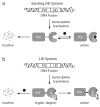
Figure 1
Figure 1
Two strategies for ligand-regulated protein stability. (a) Schematic representation of the destabilizing domain (DD) technology. Addition of ligand stabilizes the protein-of-interest (POI). (b) Schematic illustration of the ligand-induced degradation (LID) technology in which addition of the ligand destabilizes rather than stabilizes its target.
Figure 2
Figure 2
. Characterization of the LID domain. (a) NIH3T3 cells stably expressing YFP-LID were treated with either vehicle or 10 μM MG132 in the presence or absence of 1 μM Shield-1 for 6 h followed by flow cytometry analysis. Lysates from the cells described were resolved by SDS-PAGE and immunoblotted with either anti-YFP or anti-Hsp90 antibodies (Supplementary Fig. 9). (b) Fluorescence microscopy of cells stably expressing the YFP-LID fusion. Cells were treated with either vehicle or 1 μM Shield-1 for 24 h and analyzed using epifluorescence microscopy. HcRed serves as a marker for infection. Insert scalebars represent 10 μm. (c) Cells stably expressing the YFP-LID fusion were treated with various concentrations of Shield-1 (1 μM to 1 pM) and monitored by flow cytometry. (d) The degradation (squares) of YFP-LID was monitored at various times following addition of Shield-1. The recovery (triangles) of YFP-LID was monitored at various time points after depletion of Shield-1 from the culture media. Both experiments were analyzed by flow cytometry. The maximum observed fluorescence intensity for each construct was set to 100%. (e) Cells stably expressing either the YFP-LID or the YFP-19mer fusion proteins were treated with vehicle (−) or with 1 μM Shield-1 for 24 h (+) and analyzed by flow cytometry. The error bars represent the s.d. of the mean based on at least two experiments.
Figure 3
Figure 3
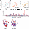
. NMR analysis of FKBP and the LID domain. 1H/15N HSQC spectra of (a) FKBP (red) compared to LID (cyan), (b) FKBP (red) compared to FKBP bound to Shield-1 (orange), and (c) FKBP bound to Shield-1 (orange) compared to LID bound to Shield-1 (blue). (d) Relative chemical shift perturbations of FKBP compared to LID (blue bars) as well as FKBP compared to the FKBP•Shield-1 complex (red bars). Residues that could not be assigned in the HSQC-spectra are indicated with negative bars. (e) Chemical shift perturbations mapped onto the structure of FKBP complexed with a Shield-1 analog (PDB:1BL4). Residues experiencing significant chemical shift perturbations (δavgmax > 0.2) are colored red, moderate perturbations are colored purple (0.1 < δavgmax < 0.2), and minor perturbations are shown in blue (δavgmax <0.1).
Figure 4
Figure 4
Characterization of the 19-residue peptide degron. (a) Alanine was substituted at seventeen positions of the 19-residue peptide fused to YFP-FKBP(F36V). Cells were stably transduced with the appropriate retrovirus and after 24 hours were treated with vehicle or 2 μM Shield-1 for 24 hours and YFP levels were scored by analytical flow cytometry. (b) Four C-terminal truncation mutants of the YFP-LID domain were prepared, and NIH3T3 cells stably transduced with these constructs were treated with either vehicle or 2 μM Shield-1 for 24 hours and evaluated by flow cytometry. Deleted residues are indicated as asterisks. (c) The 19-residue peptide and deletion mutants of this degron were fused directly to YFP to evaluate the strength of these degrons. Deleted residues are indicated as asterisks. The error bars represent the s.d. of the mean based on at least two experiments.
Figure 5
Figure 5
LID domain regulates transcription factors. (a) The LID domain was fused to the C-termini of six transcription factors that were independently transduced into NIH3T3 cells. Cell populations were treated with either vehicle or 2 μM Shield-1, and cell lysates were immunoblotted with antibodies against the indicated proteins (Oct4, Klf4), anti-HA (Sox2, Myc, Lin28) or anti-Flag (Nanog). α-Tubulin serves as the loading control (LC). Lane A represents untransduced NIH3T3 cells. Lanes B and C are from cells that were treated with vehicle (−) or with 2 μM Shield-1 for 24 h (+). Full western blots are shown in Supplementary Fig. 10. (b) Nuclear reprogramming of MEFs derived from transgenic mice encoding an Oct4 promoter driving GFP. MEF(Oct4/GFP) cells (ref. 24) were transduced with Sox2, Myc, Klf4, and Oct4 (left) or with Sox2, Myc, Klf4, and Oct4-LID (right). Cells were treated with vehicle or with 2 μM Shield-1 on day 3 post-infection. Alkaline phosphatase (AP) staining was performed on day 14 post-infection. Insert scalebars represent 5 mm. (c) GFP positive cells were transferred on day 19 to a new feeder layer. APC conjugated SSEA-1 (Stage-Specific Embryonic Antigen-1) antibody staining was performed on day 24 post-infection. Insert scalebars represent 50 μm.
Figure 6
Figure 6
Shield-1 can simultaneously stabilize and destabilize specific targets. NIH3T3 cells stably expressing both YFP-LID and DD-mCherry were treated with various concentrations of Shield-1 for 24 h, and the fluorescent signals in the appropriate channels were monitored by flow cytometry (a) and fluorescence microscopy (b). Insert scalebars represent 10 μm.

Comment in

References

    1. Furth PA, et al. Temporal control of gene expression in transgenic mice by a tetracycline-responsive promoter. Proc. Natl. Acad. Sci. U. S. A. 1994;91:9302–9306. - PMC - PubMed
    1. Ryding ADS, Sharp MGF, Mullins JJ. Conditional transgenic technologies. J. Endocrinol. 2001;171:1–14. - PubMed
    1. Banaszynski LA, Wandless TJ. Conditional control of protein function. Chem. Biol. 2006;13:11–21. - PubMed
    1. Raina K, Crews CM. Chemical Inducers of Targeted Protein Degradation. J. Biol. Chem. 2010;285:11057–11060. - PMC - PubMed
    1. Zhou P, Bogacki R, McReynolds L, Howley PM. Harnessing the ubiquitination machinery to target the degradation of specific cellular proteins. Mol. Cell. 2000;6:751–756. - PubMed

Publication types