. 2011 Jul 3;14(8):973-83.
doi: 10.1038/nn.2857.

A CaMKIIβ signaling pathway at the centrosome regulates dendrite patterning in the brain

Affiliations

A CaMKIIβ signaling pathway at the centrosome regulates dendrite patterning in the brain

Sidharth V Puram et al. Nat Neurosci. .

Abstract

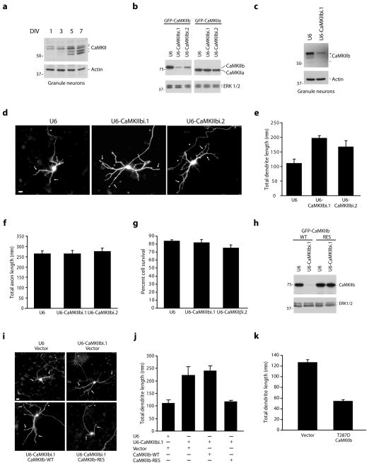
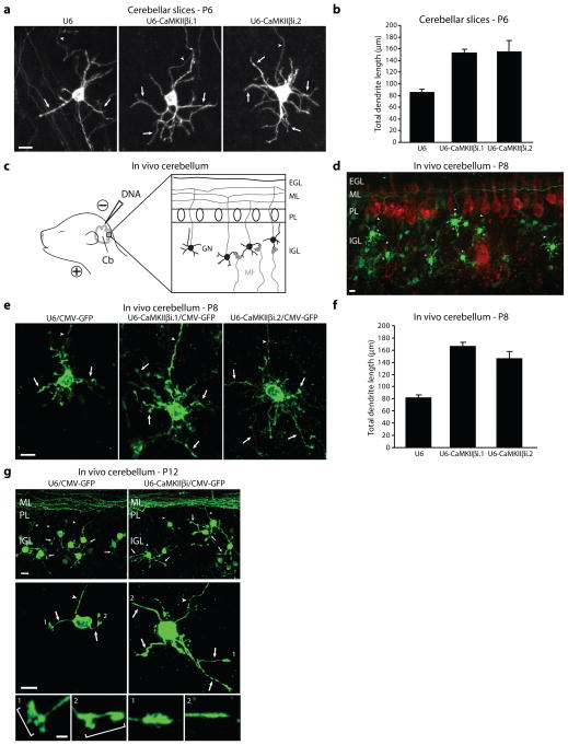
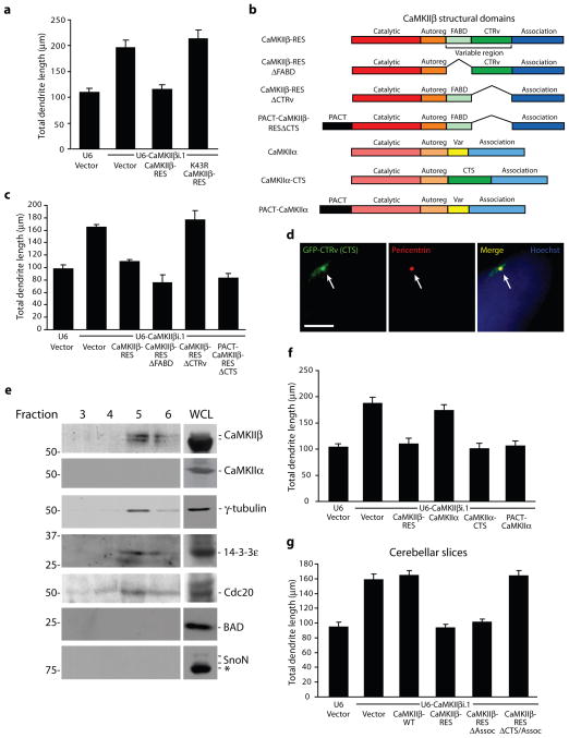
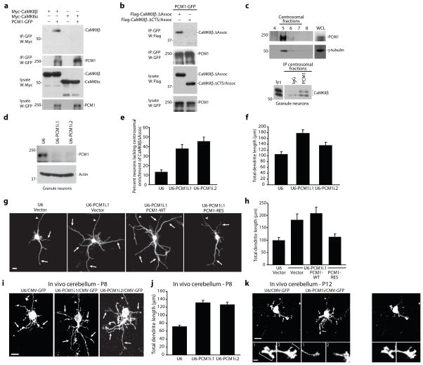
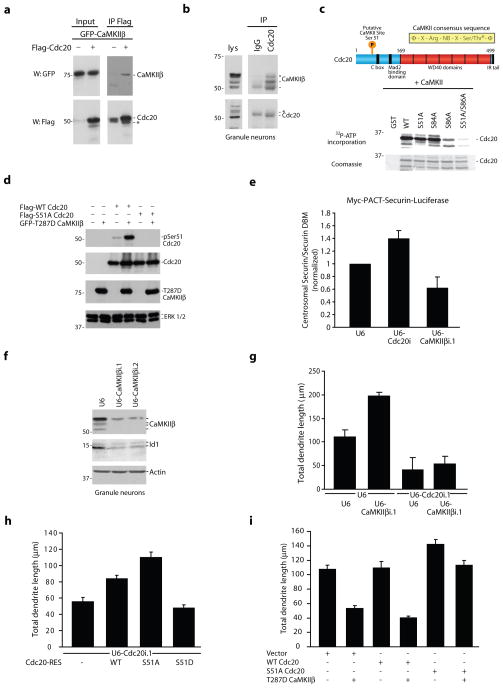
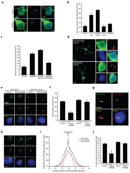
The protein kinase calcium/calmodulin-dependent kinase II (CaMKII) predominantly consists of the α and β isoforms in the brain. Although CaMKIIα functions have been elucidated, the isoform-specific catalytic functions of CaMKIIβ have remained unknown. Using knockdown analyses in primary rat neurons and in the rat cerebellar cortex in vivo, we report that CaMKIIβ operates at the centrosome in a CaMKIIα-independent manner to drive dendrite retraction and pruning. We also find that the targeting protein PCM1 (pericentriolar material 1) localizes CaMKIIβ to the centrosome. Finally, we uncover the E3 ubiquitin ligase Cdc20-APC (cell division cycle 20-anaphase promoting complex) as a centrosomal substrate of CaMKIIβ. CaMKIIβ phosphorylates Cdc20 at Ser51, which induces Cdc20 dispersion from the centrosome, thereby inhibiting centrosomal Cdc20-APC activity and triggering the transition from growth to retraction of dendrites. Our findings define a new, isoform-specific function for CaMKIIβ that regulates ubiquitin signaling at the centrosome and thereby orchestrates dendrite patterning, with important implications for neuronal connectivity in the brain.


Figures

Figure 1
Figure 1. CaMKIIβ restricts the elaboration of dendrites
a, Lysates of granule neurons were immunoblotted with the indicated antibodies. DIV = days in vitro. Full-length blots for all immunoblotting analyses are presented in Supplementary Fig. 11. b, Lysates of COS cells transfected with GFP-CaMKIIβ or GFP-CaMKIIα together with the CaMKIIβ RNAi or control U6 plasmid were immunoblotted with the indicated antibodies. c, Lysates of granule neurons electroporated with the CaMKIIβ RNAi or control U6 plasmid were immunoblotted with the indicated antibodies. d, Granule neurons transfected with a CaMKIIβ RNAi or control U6 plasmid together with GFP were subjected to immunocytochemistry using the GFP antibody. In all images of neuronal morphology, arrows and arrowheads indicate dendrites and axons, respectively. Scale bar = 10 μm. e, Total dendrite length for granule neurons treated as in (d) was quantified and is presented as mean + SEM. Total dendrite length was significantly increased in CaMKIIβ knockdown neurons compared to control U6-transfected neurons (ANOVA; p < 0.005). 270 neurons were measured. f, Granule neurons were analyzed as in (d). Total axon length was not significantly different in CaMKIIβ knockdown neurons and control U6-transfected neurons. g, Granule neurons were transfected with a CaMKIIβ RNAi or control U6 plasmid and analyzed for cell survival. Cell survival was not significantly different in CaMKIIβ knockdown neurons and control U6-transfected neurons. h, Lysates of COS cells transfected with GFP-CaMKIIβ-WT or GFP-CaMKIIβ-RES together with the CaMKIIβ RNAi or control U6 plasmid were immunoblotted with the indicated antibodies. i, Granule neurons transfected with the CaMKIIβ RNAi or control U6 plasmid together with the expression plasmid encoding CaMKIIβ-WT, CaMKIIβ-RES, or control vector and GFP were analyzed as in (d). Scale bar = 10 μm. j, Total dendrite length for granule neurons treated as in (i) was quantified. CaMKIIβ-RES, but not CaMKIIβ-WT, significantly reduced total dendrite length compared to control vector in the background of CaMKIIβ RNAi (ANOVA; p < 0.005). 360 neurons were measured. k, Granule neurons transfected with constitutively active T287D CaMKIIβ or control vector were analyzed as in (d). Total dendrite length was significantly reduced in T287D CaMKIIβ-expressing neurons compared to control vector-transfected neurons (t-test, p < 0.0005). 180 neurons were measured.
Figure 2
Figure 2. CaMKIIβ regulates dendrite patterning in vivo
a, Post-natal day six (P6) rat cerebellar slices transfected by a biolistics method with the CaMKIIβ RNAi or control U6 plasmid together with GFP were subjected to immunohistochemistry using the GFP antibody. Scale bar = 10 μm. b, CaMKIIβ knockdown neurons had significantly longer dendrites compared to control U6-transfected neurons (ANOVA, p < 0.0001). 199 neurons were measured. c, Schematic of in vivo electroporation approach. d, Rat pups electroporated in vivo with a U6-CaMKIIβi/CMV-GFP RNAi or control U6/CMV-GFP plasmid were sacrificed five days after electroporation and cerebella were subjected to immunohistochemistry using the GFP and Calbindin antibody. Representative control-transfected granule neurons are shown. Scale bar = 10 μm. e, Rat pups were electroporated as in (d) and representative neurons for each condition are shown. Scale bar = 10 μm. f, IGL granule neurons analyzed as in (d) were subjected to morphometric analysis. Total dendrite length was significantly increased in IGL granule neurons in CaMKIIβ knockdown animals compared to control U6 animals (ANOVA, p < 0.0001). 266 neurons were measured. g, Rat pups electroporated in vivo with the U6-CaMKIIβi/CMV-GFP RNAi or control U6/CMV-GFP plasmid were sacrificed nine days after electroporation and cerebella were analyzed as in (d). (Top) Representative cerebellar sections from each condition are shown. Scale bar = 10 μm. (Bottom) Representative IGL granule neurons for each condition are shown. Scale bar = 10 μm. Inset: Zoomed view of dendritic tips of individual neurons. Scale bar = 2.5 μm. Bracket identifies dendritic claws.
Figure 3
Figure 3. CaMKIIβ operates at the centrosome to specifically control dendrite patterning
a, Granule neurons transfected with the CaMKIIβ RNAi or control U6 plasmid together with CaMKIIβ-RES, K43R CaMKIIβ-RES, or control vector and GFP were analyzed as in Fig. 1d. CaMKIIβ-RES, but not K43R CaMKIIβ-RES, significantly reduced total dendrite length compared to control vector in the background of CaMKIIβ RNAi (ANOVA; p < 0.0001). 390 neurons were measured. b, Schematic of major structural domains of CaMKIIβ. c, Granule neurons transfected with the CaMKIIβ RNAi or control U6 plasmid together with CaMKIIβ-RES, CaMKIIβ-RESΔFABD, CaMKIIβ-RESΔCTRv, PACT-CaMKIIβ-RESΔCTS, or control vector and GFP were analyzed as in (a). CaMKIIβ-RES, CaMKIIβ-RESΔFABD, and PACT-CaMKIIβ-RESΔCTS, but not CaMKIIβ-RESΔCTRv, significantly reduced total dendrite length compared to control vector in the background of CaMKIIβ RNAi (ANOVA; p < 0.0001). 660 neurons were measured. d, Granule neurons transfected with GFP fused to the C-terminal variable region (GFP-CTRv) were subjected to immunocytochemistry using the GFP and pericentrin antibody. Arrows indicate co-localization of GFP-CTRv with pericentrin. Scale bar = 5 μm. e, Fractions isolated from a granule neuron centrosome preparation were immunoblotted using the indicated antibodies. Asterisk indicates non-specific band. WCL, whole cell lysate. f, CaMKIIβ-RES, CaMKIIα-CTS, and PACT-CaMKIIα, but not CaMKIIα, significantly reduced total dendrite length compared to control vector in the background of CaMKIIβ RNAi in granule neurons (ANOVA; p < 0.0001). 600 neurons were measured. g, CaMKIIβ-RES and CaMKIIβ-RESΔAssoc, but not CaMKIIβ-WT or CaMKIIβ-RESΔAssoc/CTS, significantly reduced granule neuron total dendrite length compared to control vector in the background of CaMKIIβ RNAi in cerebellar slices (ANOVA; p < 0.0001). 479 neurons were measured.
Figure 4
Figure 4. The centrosomal targeting protein PCM1 localizes CaMKIIβ at the centrosome
a, Lysates of 293T cells transfected with Myc-CaMKIIβ or Myc-CaMKIIα together with PCM1-GFP or control vector were immunoprecipitated using the GFP antibody and immunoblotted with the indicated antibodies. b, Lysates of 293T cells transfected with Flag-CaMKIIβΔAssoc or Flag-CaMKIIβΔCTS/Assoc together with PCM1-GFP were immunoprecipitated using the GFP antibody and immunoblotted with the Flag or GFP antibody. c, Fractions isolated from a granule neuron centrosome preparation were immunoblotted using the PCM1 or γ-tubulin antibody. Pooled centrosomal fractions were immunoprecipitated with the PCM1 or control (IgG) antibody and immunoblotted with the CaMKIIβ antibody. d, Lysates of granule neurons electroporated with the PCM1 RNAi or control U6 plasmid were immunoblotted with the indicated antibodies. e, Granule neurons transfected with a PCM1 RNAi or control U6 plasmid together with GFP-CaMKIIβΔAssoc were analyzed as in Fig. 3d. The percentage of neurons lacking centrosomal enrichment of GFP-CaMKIIβΔAssoc was significantly increased in PCM1 knockdown neurons compared to control U6-transfected neurons (ANOVA, p < 0.005). 394 neurons were analyzed. f, Total dendrite length was significantly increased in PCM1 knockdown neurons compared to control U6-transfected neurons (ANOVA; p < 0.0001). 180 neurons were measured. g, Granule neurons transfected with the PCM1 RNAi or control U6 plasmid together with PCM1-WT, PCM1-RES, or control vector and GFP were analyzed as in (f). Scale bar = 10 μm. h, PCM1-RES, but not PCM1-WT, significantly reduced total dendrite length compared to control vector in the background of PCM1 RNAi (ANOVA; p < 0.0001). 240 neurons were measured. i, Rat pups electroporated in vivo with a U6-PCM1i/CMV-GFP RNAi or control U6/CMV-GFP plasmid were sacrificed at P8 and analyzed as in Fig. 2d. Scale bar = 10 μm. j, Total dendrite length was significantly increased in IGL granule neurons in PCM1 knockdown animals compared to control U6 animals (ANOVA, p < 0.0001). 270 neurons were measured. k, Rat pups electroporated in vivo with the U6-PCM1i/CMV-GFP RNAi or control U6/CMV-GFP plasmid were sacrificed at P12 and analyzed as in Fig. 2g. Scale bar = 10 μm. Inset: Scale bar = 2.5 μm.
Figure 5
Figure 5. CaMKIIβ phosphorylates Cdc20 at Ser51 and thereby inhibits centrosomal Cdc20-APC activity in neurons
a, Lysates of 293T cells transfected with GFP-CaMKIIβ together with Flag-Cdc20 or control vector were immunoprecipitated using the Flag antibody and immunoblotted with the indicated antibodies. b, Lysates of granule neurons were immunoprecipitated with the Cdc20 or control (IgG) antibody and immunoblotted with the indicated antibodies. Asterisk indicates IgG heavy chain. c, (Top) Schematic of CaMKII consensus sequence. (Bottom) Recombinant WT and S51A, S84A, S86A, and S51A/S86A mutants of an N-terminal region of Cdc20 (1–101) fused to GST were incubated in vitro with purified CaMKII. d, Lysates of 293T cells transfected with Flag-WT Cdc20 or Flag-S51A Cdc20 together with GFP-T287D CaMKIIβ or control vector were immunoblotted with the indicated antibodies. e, Granule neurons were transfected with the Cdc20 RNAi, CaMKIIβ RNAi, or control U6 plasmid together with Myc-PACT-securin-luciferase or Myc-PACT-securin-DBM-luciferase and analyzed by luminometry. The relative amount of the centrosomal securin-luciferase reporter was significantly increased in Cdc20 knockdown neurons and significantly reduced in CaMKIIβ knockdown neurons compared to control U6-transfected neurons (Kruskal-Wallis; p < 0.01, n = 4). f, Lysates of granule neurons electroporated with the CaMKIIβ RNAi or control U6 plasmid were immunoblotted with the indicated antibodies. g, CaMKIIβ knockdown significantly increased total dendrite length compared to control, and Cdc20 RNAi significantly reduced total dendrite length in granule neurons in the presence or absence of CaMKIIβ RNAi (ANOVA, p < 0.0001). 420 neurons were measured. h, Expression of S51A Cdc20-RES, but not S51D Cdc20-RES, significantly increased total dendrite length compared to control vector or WT Cdc20-RES in the background of Cdc20 RNAi in granule neurons (ANOVA, p < 0.0001). 428 neurons were measured. i, Expression of T287D CaMKIIβ significantly reduced total dendrite length compared to control in the background of control vector or WT Cdc20, but not in the background of S51A Cdc20 (ANOVA, p < 0.0001). 540 neurons were measured.
Figure 6
Figure 6. CaMKIIβ-induced phosphorylation of Cdc20 promotes Cdc20 dispersion from the centrosome, leading to the restriction of dendrite elaboration
a, Granule neurons transfected with CaMKIIβ or control vector together with farnesylated GFP (fGFP) were subjected to immunocytochemistry using the GFP or Cdc20 antibody. Arrows indicate Cdc20 localized to the centrosome, while arrowheads denote dispersed Cdc20. Scale bar = 5 μm. b, The percentage of neurons displaying dispersed endogenous Cdc20 was significantly increased in T287D CaMKIIβ- and WT CaMKIIβ-expressing neurons, but not K43R CaMKIIβ- or CaMKIIα-expressing neurons, as compared to control vector-transfected neurons (ANOVA; p < 0.0001). 622 neurons were analyzed. c, The percentage of neurons displaying dispersed endogenous Cdc20 was significantly increased in CaMKIIβ- and CaMKIIβΔAssoc-expressing neurons, but not CaMKIIβΔCTS/Assoc-expressing neurons, as compared to control vector-transfected neurons (ANOVA; p < 0.0001). 360 neurons were analyzed. d, Granule neurons transfected with T287D CaMKIIβ together with fGFP were analyzed as in (a). Arrows indicate dendrites, while dashed box indicates the magnified region in inset images. Scale bar = 5 μm. e, Granule neurons transfected with GFP-Cdc20 or GFP-PACT-Cdc20 together with T287D CaMKIIβ or control vector were analyzed as in Fig. 3d. Arrows indicate the location of the centrosome, which is labeled with pericentrin. Scale bar = 5 μm. f, Total dendrite length was significantly reduced in T287D CaMKIIβ-expressing neurons compared to control-transfected neurons in the background of Cdc20 (ANOVA, p < 0.0005), but not in the background of PACT-Cdc20. 360 neurons were measured. g, Granule neurons transfected with GFP-centrin were subjected to immunocytochemistry using the GFP or phosphoSer51-Cdc20 antibody. Arrowheads indicate phosphoSer51-Cdc20 immunoreactivity. Scale bar = 5 μm. h, Granule neurons transfected with GFP-Cdc20, GFP-S51A Cdc20, or GFP-S51D Cdc20 were analyzed as in (e). Scale bar = 5 μm. i, Granule neurons treated as in (h) were analyzed by linescan analysis. The location of the centrosome was identified based on pericentrin immunoreactivity. S51A Cdc20 had enhanced signal at the centrosome and reduced cytosolic signal compared to WT Cdc20. In contrast, S51D Cdc20 had reduced centrosomal signal and broader non-centrosomal signal compared to WT Cdc20 (based on peak centrosomal signal intensity, ANOVA, p < 0.0001). 270 neurons were analyzed. j, Expression of T287D CaMKIIβ significantly reduced the percentage of neurons with centrosomally-enriched WT Cdc20 compared to control vector, but had no effect on the centrosomal localization of S51A Cdc20 (ANOVA; p < 0.01). 361 neurons were analyzed.

Comment in

References

    1. Altman J, Bayer S. Development of the Cerebellar System: In Relation to Its Evolution, Structure, and Functions. CRC Press; New York: 1997.
    1. Palay S, Chan-Palay V. Cerebellar Cortex: Cytology and Organization. Springer-Verlag; New York: 1974.
    1. Lee A, et al. Control of dendritic development by the Drosophila fragile X-related gene involves the small GTPase Rac1. Development. 2003;130:5543–5552. - PubMed
    1. Kaufmann WE, Moser HW. Dendritic anomalies in disorders associated with mental retardation. Cereb Cortex. 2000;10:981–991. - PubMed
    1. Corty MM, Matthews BJ, Grueber WB. Molecules and mechanisms of dendrite development in Drosophila. Development. 2009;136:1049–1061. - PMC - PubMed

Publication types

MeSH terms