. 2012 Feb 9;31(6):739-51.
doi: 10.1038/onc.2011.276. Epub 2011 Jul 4.

Regulated ATF5 loss-of-function in adult mice blocks formation and causes regression/eradication of gliomas

Affiliations
Free PMC article

Regulated ATF5 loss-of-function in adult mice blocks formation and causes regression/eradication of gliomas

A Arias et al. Oncogene. .
Free PMC article

Abstract

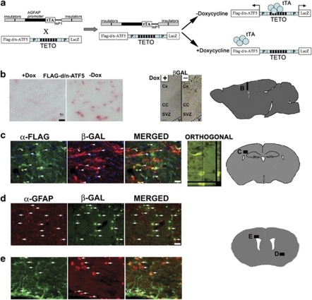
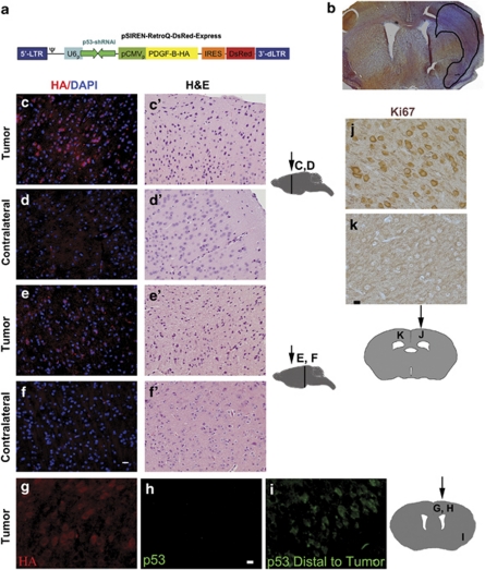
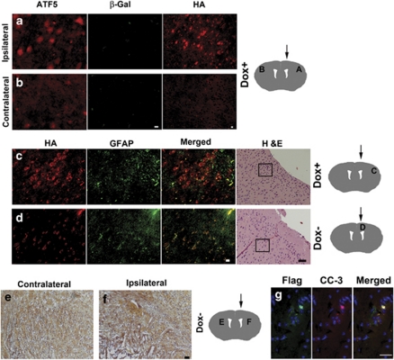
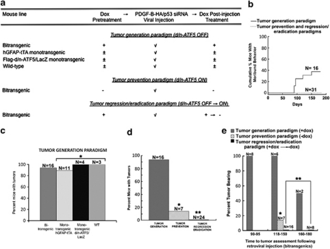
Glioblastomas are among the most incurable cancers. Our past findings indicated that glioblastoma cells, but not neurons or glia, require the transcription factor ATF5 (activating transcription factor 5) for survival. However, it was unknown whether interference with ATF5 function can prevent or promote regression/eradication of malignant gliomas in vivo. To address this issue, we created a mouse model by crossing a human glial fibrillary acidic protein (GFAP) promoter-tetracycline transactivator mouse line with tetracycline operon-dominant negative-ATF5 (d/n-ATF5) mice to establish bi-transgenic mice. In this model, d/n-ATF5 expression is controlled by doxycycline and the promoter for GFAP, a marker for stem/progenitor cells as well as gliomas. Endogenous gliomas were produced with high efficiency by retroviral delivery of platelet-derived growth factor (PDGF)-B and p53-short hairpin RNA (shRNA) in adult bi-transgenic mice in which expression of d/n-ATF5 was spatially and temporally regulated. Induction of d/n-ATF5 before delivery of PDGF-B/p53-shRNA virus greatly reduced the proportion of mice that formed tumors. Moreover, d/n-ATF5 induction after tumor formation led to regression/eradication of detectable gliomas without evident damage to normal brain cells in all 24 mice assessed.


Figures

Figure 1
Figure 1
Cells within the neurogenic zones of postnatal mouse brain express ATF5. A paraffin-embedded 28-day postnatal mouse brain was sectioned and immunostained for the following markers: (a) ATF5 (brown). (b) ATF5 (brown) and GFAP (red). The × 2.33 magnification inset shows a subset of cells co-stained for GFAP and ATF5. (c) ATF5 (brown) and GFAP (red). Arrows show a subset of cells along the ventricle and corpus callosum (CC) that co-stain for ATF5 and GFAP. (d) Nestin. (e) Musashi-1. In (a)–(e), note staining within the ependymal zone (EZ), SVZ, CC and the junction between the corpus callosum and the striatum (S). (f) ATF5 (brown) and GFAP (red) in the subgranular zone (SGZ) of the hippocampal dentate gyrus. Arrows show cells co-stained for ATF5 and GFAP. Scale bar=20 μm. Diagrams show locations in the brain of corresponding panel letter (adapted from http://www.hms.harvard.edu/research/brain/atlas).
Figure 2
Figure 2
Regulated expression of d/n-ATF5 in bi-transgenic hGFAP-tTA × pBi-TETO-3-Flag-d/n-ATF5/β-gal mice. (a) Scheme for generation and utilization of bi-transgenic hGFAP-tTA × pBi-TETO-3-Flag-d/n-ATF5/β-gal mice. The hGFAP promoter drives the tTA, and the tTA binds to TETO in the absence of doxycycline (Dox) to induce bi-directional co-expression of the Flag-d/n-ATF5 and β-gal (LacZ) transgenes. (b) Regulated expression of Flag-d/n-ATF5 (left-hand panels) and β-gal (right-hand panels) in the cortex, SVZ and CC of bi-transgenic mice. One bi-transgenic mouse was maintained on Dox at conception and killed 63 days after birth (+Dox), whereas the other was maintained on Dox from conception until 35 days after birth, and then withdrawn from the drug and killed 28 days later (−Dox). Colorimetric staining of Flag-tagged d/n-ATF5 in the cortex (sagittal section) was achieved with anti-FLAG M2 antibody and Vulcan Fast Red. Scale bar=10 μm. X-gal substrate was used for β-gal colorimetric detection. V=lateral ventricle; CC=corpus callosum; Cx=cortex; SVZ=subventricular zone. Scale bar=20 μm. (c) Coordinately regulated expression of Flag-d/n-ATF5 and β-gal within the SVZ and near the CA1 region of the hippocampus (orthogonal view) in a 79-day-old bi-transgenic mouse raised without Dox since conception. Immunofluorescence staining was carried out with anti-Flag (for detection of Flag-d/n-ATF5; green) and β-gal (red). Arrows in the merged panel indicate cell colocalization. The confocal (2 μm) orthogonal section image shows the xy and yz location of both β-gal and Flag-d/n-ATF5 within the same cell. (d) Regulated expression of β-gal in GFAP+ cells within the cortex of a bi-transgenic mouse. The animal was maintained on Dox from conception until 204 days after birth and the drug was then withdrawn until being killed 50 days later. Panels show immunostaining with anti-GFAP (red) and anti-β-gal (green) and arrows show the locations of cells in which both are co-expressed. (e) Regulated expression of β-gal in GFAP+ cells within the SVZ of a bi-transgenic mouse. The mouse was maintained on doxycycline from conception until 167 days after birth and the drug was then withdrawn until being killed 42 days later. Panels show immunostaining with anti-GFAP (green) and anti-β-gal (red) and arrows show the locations of cells in which both are co-expressed. Diagrams show the brain areas where photographs were taken (adapted from BrainMaps.org and http://www.hms.harvard.edu/research/brain/atlas). Scale bar=20 μm (ce).
Figure 3
Figure 3
Retrovirus encoding PDGF-B-HA and p53-short hairpin RNA (shRNA) induces gliomas within mouse brains, and knocks down p53. (a) Scheme of self-inactivating retroviral construct used to induce gliomas. The 5′-long terminal repeat is intact, but the 3′-deleted long terminal repeat possesses a deletion to prevent self-replication. The U6 pol III promoter drives transcription of p53-shRNA, whereas the cytomegalovirus promoter drives transcription of the bicistronic PDGF-B-HA-IRES-DsRed-Express reporter. (b) Frozen section. H&E staining reveals a glioma induced in a doxycycline-treated hGFAP-tTA × pBi-TETO-3-Flag-d/n-ATF5/β-gal bi-transgenic mouse 100 days after stereotactic injection of retrovirus expressing PDGF-B-HA and p53-shRNA. Area of glioma is enclosed within black line. (c, c′–f, f′) Tumor cells induced by PDGF-B-HA/p53-shRNA express the HA tag and have dark hyperchromatic nuclei. The bi-transgenic mouse was continuously maintained with doxycycline from conception and was killed 135 days after injection with PDGF-B-HA/p53-shRNA retrovirus. Paraffin-embedded brain sections of PDGF-B-HA/p53-shRNA were immunostained with rabbit anti-HA antibody (red) and 4′,6-diamidino-2-phenylindole (blue) (cf) or H&E (c′–f′). c, c′, e and e′ show PDGF-B-HA/p53-shRNA-induced tumors, whereas d, d′, f and f′ are from the corresponding tumor-free contralateral hemisphere. Scale bar=12 μm. (gi) Tumors induced by PDGF-B-HA/p53-shRNA express the HA tag, but not p53. Photos show immunostained (for HA and p53) paraffin-embedded brain sections from a bi-transgenic mouse continuously exposed to doxycycline from conception and killed 135 days after retroviral injection. The HA+, p53 sections in (g) and (h) include tumor tissue, whereas the p53+ (green) section in (i) was outside the tumor margins. Scale bar=5 μm. (j, k) Gliomas induced by PDGF-B-HA/p53-shRNA have a proliferative index of 100% calculated from Ki67 immunostaining. A monotransgenic hGFAP-tTA mouse was continuously fed with doxycycline from conception and killed 31 days after stereotactic injection with PDGF-B-HA/p53-shRNA retrovirus. Paraffin-embedded brain sections were immunostained for Ki67 antiserum with visualization by immunoperoxidase (brown). (j) Ki67+ cells in a glioma induced in the injected hemisphere, whereas (k) shows the absence of Ki67 staining in normal tissue in the contralateral hemisphere. Scale bar=10 μm. Diagrams of brain sections in (c)–(k) show sites of retroviral injection (arrows) and corresponding areas where photographs were taken (vertical lines and letters). Diagrams are adapted from http://www.hms.harvard.edu/research/brain/atlas.
Figure 4
Figure 4
Bi-transgenic hGFAP-tTA × pBi-TETO-3-Flag-d/n-ATF5/β-gal mice do not harbor detectable gliomas after induction of d/n-ATF5. (a, b) PDGF-B-HA/p53-shRNA-induced glioma in a bi-transgenic mouse expresses ATF5, but not the d/n-ATF5/β-gal transgene. The mouse was continuously maintained on doxycycline from conception to suppress d/n-ATF5 induction and was killed 135 days post-stereotactic injection with PDGF-B-HA/p53-shRNA retrovirus. Brain sections were stained with rabbit anti-ATF5 antibody (red) and chicken anti-β-gal antibody (green). Note strong immunostaining for ATF5 and lack of β-gal immunostaining in tumor (a) and only weak scattered, low-level ATF5 immunoreactivity in the contralateral hemisphere (b). H&E staining for (a) is from the same brain shown in Figure 3c′, and H&E staining for (b) is from the same brain shown in Figure 3d′. Scale bar=6 μm. (c, d) Immunostaining for the HA tag (red) and GFAP (green) detects a glioma, as shown by H&E staining, in a mouse in which expression of d/n-ATF5 was suppressed (c), but not in a mouse in which d/n-ATF5 was induced (d). The right-hand panels show H&E staining to confirm the presence or absence of a glioma. Boxes within the H&E photos show where the anti-HA and anti-GFAP immunofluorescence images were taken. Scale bar=40 μm for H&E photos and 12 μm for immunofluorescence images in (c) and (d). The bi-transgenic mouse in (c) was continuously maintained on doxycycline from conception to suppress d/n-ATF5 induction and was killed 135 days post-stereotactic injection with PDGF-B-HA/p53-shRNA retrovirus. Note HA and GFAP immunostaining in tumor remote from site of injection (c). The bi-transgenic mouse in (d) was maintained on doxycycline from conception to 90 days post-injection with PDGF-B-HA/p53-shRNA retrovirus. Doxycycline was then withdrawn from the diet for 45 days to induce d/n-ATF5 and the animal was killed. Examination of serial brain sections revealed scattered HA-stained cells as shown, only at the site of viral injection. GFAP+ cells at upper right reflect astrocytic activation in response to the injection wound. Paraffin-embedded sections. (e, f) Evidence for eradication of a pre-existing tumor by induction of d/n-ATF5. The bi-transgenic mouse was maintained on doxycycline from conception to 92 days post-injection with PDGF-B-HA/p53-shRNA retrovirus. Doxycycline was then withdrawn from the diet for 50 days to induce d/n-ATF5. (e) Normal cells in contralateral hemisphere, but (f) lesions in the neutrophil remote from the viral injection site in the ipsilateral hemisphere. Scale bar=20 μm. (g) Co-expression of cleaved caspase-3 (CC-3) and d/n-ATF5 in a tumor remnant in the ‘eradication/regression' paradigm. A bi-transgenic mouse was maintained on doxycycline from conception to 90 days after injection with PDGF-B-HA/p53-shRNA retrovirus. Doxycycline was then withdrawn from the diet for 50 days to induce d/n-ATF5 and the animal was killed. Brain sections that contained a remnant of a glioma were co-stained for Flag-tagged d/n-ATF5 (green) and cleaved-caspase-3 (red). Scale bar=20 μm. Diagrams show the brain areas for stereotactic injection site (arrow) with corresponding panel letter (diagrams adapted from http://www.hms.harvard.edu/research/brain/atlas).
Figure 5
Figure 5
Induction of d/n-ATF5 in pBi3-Flag-d/n-ATF5/LacZ × hGFAP-tTA bi-transgenic mice blocks formation and causes regression/eradication of gliomas induced by PDGF-B-HA/p53-shRNA. (a) Overview of experimental approach to testing the effect of d/n-ATF5 on glioma generation and regression/eradication in mice. (b) Moribund behavior in bi-transgenic mice injected with retrovirus expressing PDGF-B-HA/p53-shRNA and its prevention by induction of d/n-ATF5 in the glioma prevention and regression/eradication paradigms. Data show cumulative percentage of injected mice judged by veterinary staff to have moribund behavior as a function of time after viral injection. All mice found to exhibit moribund behavior were immediately killed and all were found to be tumor bearing. (c) Gliomas are induced with high efficiency in mice in which d/n-ATF5 is not expressed. Data show percentage of glioma formation in response to cerebral injection with PDGF-B-HA/p53-shRNA retrovirus in bi-transgenic (all treated with doxycycline from conception), monotransgenic hGFAP-tTA (three treated with doxycycline from conception, six treated with doxycycline from conception and withdrawn from the drug after viral injection, and two raised entirely without doxycycline), monotransgenic Flag-d/n-ATF5/LacZ (one treated with doxycycline from conception, one treated with doxycycline from conception and withdrawn from the drug after viral injection and two raised entirely without doxycycline) and wild-type (WT) (all treated with doxycycline from conception) mice. N indicates numbers of animals in each group. *Bi-transgenic mice versus monotransgenic mice, no statistical difference (P=1.0). (d) Induction of d/n-ATF5 suppresses generation of new gliomas and causes regression/eradication of pre-existing gliomas. Percent bi-transgenic mice with detectable gliomas is shown for the three treatment paradigms described in (a) and in the text and Materials and methods. The doxycycline-treated bi-transgenic mice in the ‘tumor generation' paradigm are those described in (b). N indicates numbers of animals in each group. *Bi-transgenic Dox+ versus bi-transgenic prevention Dox, P=0.0005; **bi-transgenic Dox+ versus bi-transgenic Dox regression/eradication, P=4 × 10⁻¹⁰. (e) Percentage of bi-transgenic mice with tumors at different times of assessment after injection with PDGF-B-HA/p53-shRNA retrovirus and subjected to the three treatment paradigms described in (a). Between 118 and 180 days. *Bi-transgenic Dox+ versus bi-transgenic prevention Dox, P=0.01; **bi-transgenic Dox+ versus bi-transgenic Dox regression/eradication, P=0.000002. Animals are those described in panels c and d. N indicates the number of animals assessed in each group at each time.

References

    1. Alvarez-Buylla A, Lim DA. For the long run: maintaining germinal niches in the adult brain. Neuron. 2004;41:683–686. - PubMed
    1. Angelastro JM, Canoll PD, Kuo J, Weicker M, Costa A, Bruce JN, et al. Selective destruction of glioblastoma cells by interference with the activity or expression of ATF5. Oncogene. 2006;25:907–916. - PubMed
    1. Angelastro JM, Ignatova TN, Kukekov VG, Steindler DA, Stengren GB, Mendelsohn C, et al. Regulated expression of ATF5 is required for the progression of neural progenitor cells to neurons. J Neurosci. 2003;23:4590–4600. - PMC - PubMed
    1. Angelastro JM, Mason JL, Ignatova TN, Kukekov VG, Stengren GB, Goldman JE, et al. Downregulation of activating transcription factor 5 is required for differentiation of neural progenitor cells into astrocytes. J Neurosci. 2005;25:3889–3899. - PMC - PubMed
    1. Assanah M, Lochhead R, Ogden A, Bruce J, Goldman J, Canoll P. Glial progenitors in adult white matter are driven to form malignant gliomas by platelet-derived growth factor-expressing retroviruses. J Neurosci. 2006;26:6781–6790. - PMC - PubMed

Publication types

MeSH terms

Substances