

doi: 10.1007/978-3-7091-0693-8_14.
Brain arteriovenous malformation pathogenesis: a response-to-injury paradigm
Affiliations
- PMID: 21725736
- PMCID: PMC3187860
- DOI: 10.1007/978-3-7091-0693-8_14
Item in Clipboard
Brain arteriovenous malformation pathogenesis: a response-to-injury paradigm
Acta Neurochir Suppl.
2011.
Abstract
Brain arteriovenous malformations (AVMs) are a rare but important cause of intracranial hemorrhage (ICH) in young adults. In this paper, we review both human and animal studies of brain AVM, focusing on the: (1) natural history of AVM hemorrhage, (2) genetic and expression studies of AVM susceptibility and hemorrhage, and (3) strategies for development of a brain AVM model in adult mice. These data target various mechanisms that must act in concert to regulate normal angiogenic response to injury. Based on the various lines of evidence reviewed in this paper, we propose a "response-to-injury" model of brain AVM pathogenesis.
Figures
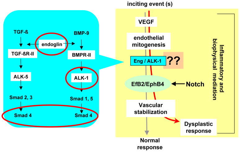
The blue shaded area is a simplified summary of presumed ALK-1 and ENG signaling via TGF-β and BMP-9 in endothelial cells (EC); the genes mutated in HHT are circled. Main components of the scheme are (a) inciting event(s) upregulate the expression of angiogenic factors, such as VEGF, which induce EC mitogenesis; newly formed vessels will develop into a stable neovasculature; (b) this process leads to a vascular dysplastic response when signaling through aberrant ALK-1 and/or ENG, or in a closely related pathway (question marks); (c) ephrinB2 and EPHB4 imbalance, possibly through involvement of Notch signaling; and (d) modifier influences, potentially genetic and/or hemodynamic. Inflammation and involvement of circulating precursor cells may be relevant.
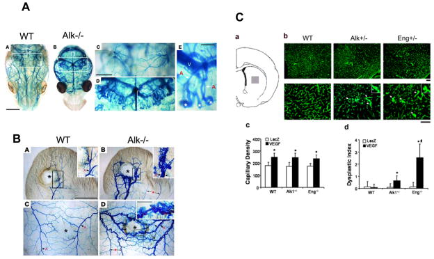
A. Endothelial Alk1 deletion results in AVMs in the brain [38]. A–E. Dissection microscopic views of vascular images of control (WT, A, C) and mutant (Alk1−/−; B, D, E) in postnatal day 3 mouse brains by latex dye injected into the left ventricle of the heart. Magnified views of blood vessels in the hipocampal area (D, E). Asterisks indicate peculiar looping of vessels at the distal tips of arteries shunting to veins (E). A, artery; V, vein. B. Wounding can induce de novo AVM formation in Alk1-deleted adult mice [38]. Vascular patterns shown by latex dye injected into the left heart of control (WT, A, C) and mutant (Alk1−/−, B, D) mice bearing wounds in the ear (A, B) or dorsal skin (C, D), 8 days after induction of Alk1 gene deletion. The images were taken after clearing in organic solvents. Center of the wound is indicated by asterisks. Note that only mutant mice developed AV shunts shown by the presence of latex dye in both arteries and veins. AV shunting and abnormal vascular morphologies were apparent only in the wound areas. Blood vessels away from the wound indicated by arrows with asterisks (B and D) showed normal appearance. Inset in D shows a magnified view of AV fistulas formed in the rim area of the mutant wound. C. Overexpression of VEGF in the striatum of Alk1 and Eng haploinsufficient mice resulted in vascular dysplasia [19]. a. Injection site (grey square). b. Angiogenic foci and dysplastic capillaries (arrows). Inserts are enlarged images of dysplastic capillaries. Scale bars: 100 μm (top panel) and 50 μm (bottom panel). c and d. Capillary density and dysplasia index. * = p<0.05, vs. AAV-LacZ group. # = p<0.05, vs. AAV-VEGF-transduced WT or Alk1+/− mice. VEGF: AAV-VEGF-injected mice; LacZ: AAV-LacZ-injected mice.
References
-
- Achrol AS, Kim H, Pawlikowska L, Poon KY, Ko NU, McCulloch CE, Zaroff JG, Johnston SC, McDermott MW, Lawton MT, Kwok PY, Young WL. Association of tumor necrosis factor-alpha-238G>A and Apolipoprotein E2 polymorphisms with intracranial hemorrhage after brain arteriovenous malformation treatment. Neurosurgery. 2007;61:731–739. - PMC - PubMed
-
- Achrol AS, Pawlikowska L, McCulloch CE, Poon KY, Ha C, Zaroff JG, Johnston SC, Lee C, Lawton MT, Sidney S, Marchuk D, Kwok PY, Young WL. Tumor necrosis factor-alpha-238G>A promoter polymorphism is associated with increased risk of new hemorrhage in the natural course of patients with brain arteriovenous malformations. Stroke. 2006;37:231–234. - PubMed
-
- Arteriovenous Malformation Study Group. Arteriovenous malformations of the brain in adults. N Engl J Med. 1999;340:1812–1818. - PubMed
Publication types
MeSH terms
Grants and funding
LinkOut - more resources
Full Text Sources
Medical