

A partially folded structure of amyloid-beta(1-40) in an aqueous environment
- PMID: 21726530
- PMCID: PMC3148408
- DOI: 10.1016/j.bbrc.2011.06.133
A partially folded structure of amyloid-beta(1-40) in an aqueous environment
Abstract
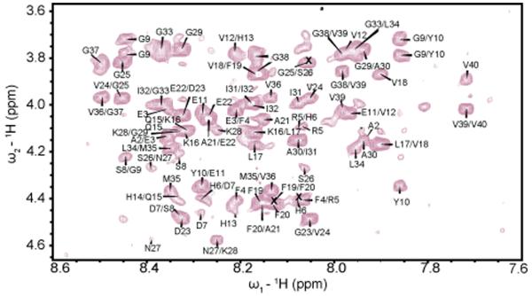
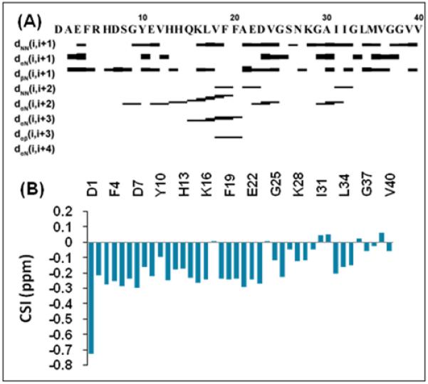
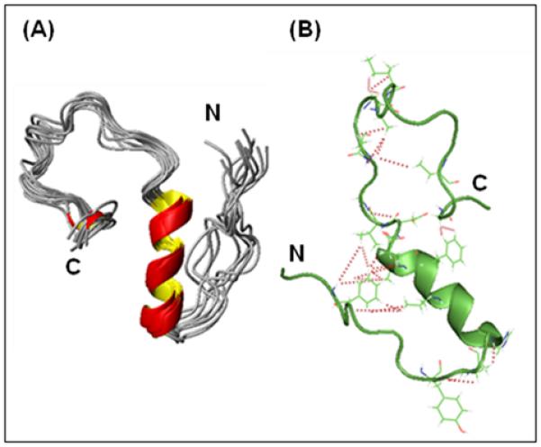
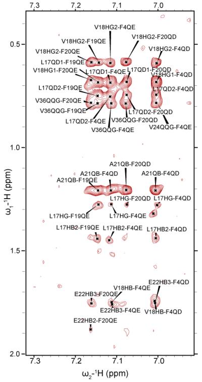
Aggregation of the Aβ(1-40) peptide is linked to the development of extracellular plaques characteristic of Alzheimer's disease. While previous studies commonly show the Aβ(1-40) is largely unstructured in solution, we show that Aβ(1-40) can adopt a compact, partially folded structure. In this structure (PDB ID: 2LFM), the central hydrophobic region of the peptide forms a 3(10) helix from H13 to D23 and the N- and C-termini collapse against the helix due to the clustering of hydrophobic residues. Helical intermediates have been predicted to be crucial on-pathway intermediates in amyloid fibrillogenesis, and the structure presented here presents a new target for investigation of early events in Aβ(1-40) fibrillogenesis.
Copyright © 2011 Elsevier Inc. All rights reserved.
Figures
References
-
- Hardy J, Allsop D. Amyloid deposition as the central event in the etiology of Alzheimer’s disease. Trends Pharmacol. Sci. 1991;12:383–388. - PubMed
-
- Smith DG, Cappai R, Barnham KJ. The redox chemistry of the Alzheimer’s disease amyloid beta peptide. Biochim. Biophys. Acta. 1768;2007:1976–1990. - PubMed
-
- Ott S, Henkel AW, Henkel MK, Redzic ZB, Kornhuber J, Wiltfang J. Pre-aggregated Aβ1-42 peptide increases tau aggregation and hyperphosphorylation after short-term application. Mol. Cell. Biochem. 2011;349:169–177. - PubMed
Publication types
MeSH terms
Substances
Grants and funding
LinkOut - more resources
Full Text Sources
Other Literature Sources
Miscellaneous
