. 2011 Sep 1;357(1):227-34.
doi: 10.1016/j.ydbio.2011.06.027. Epub 2011 Jun 25.

Shroom3 and a Pitx2-N-cadherin pathway function cooperatively to generate asymmetric cell shape changes during gut morphogenesis

Affiliations

Shroom3 and a Pitx2-N-cadherin pathway function cooperatively to generate asymmetric cell shape changes during gut morphogenesis

Timothy F Plageman Jr et al. Dev Biol. .

Abstract

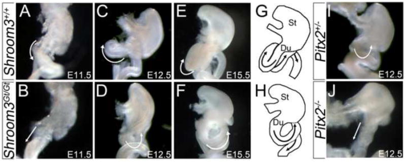
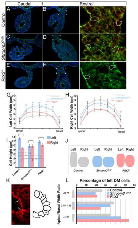
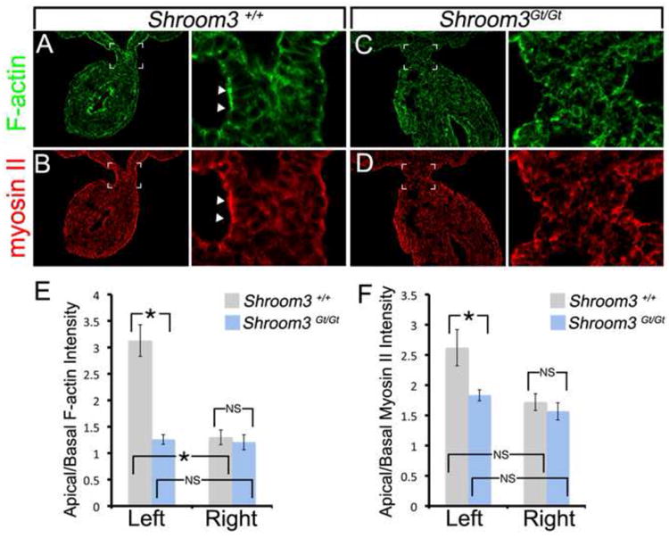
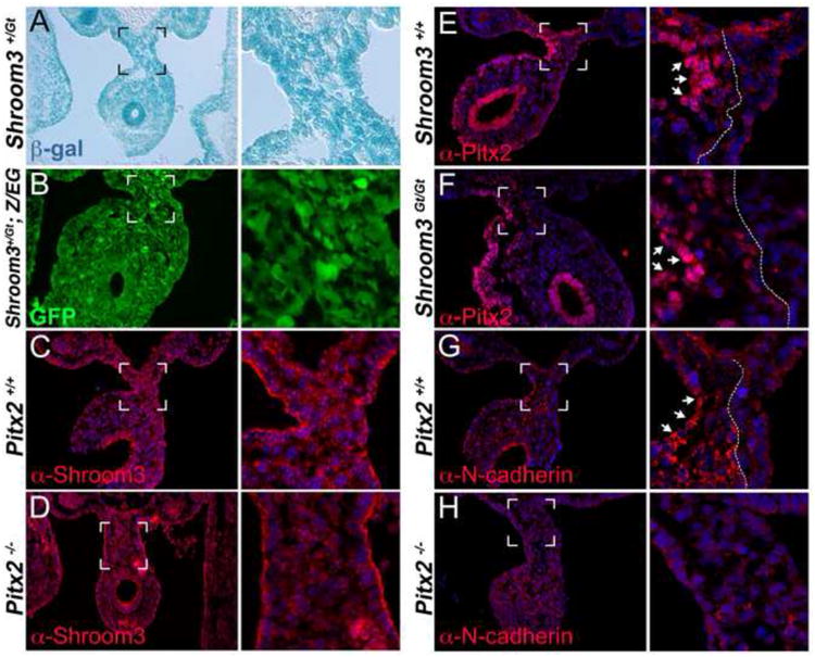
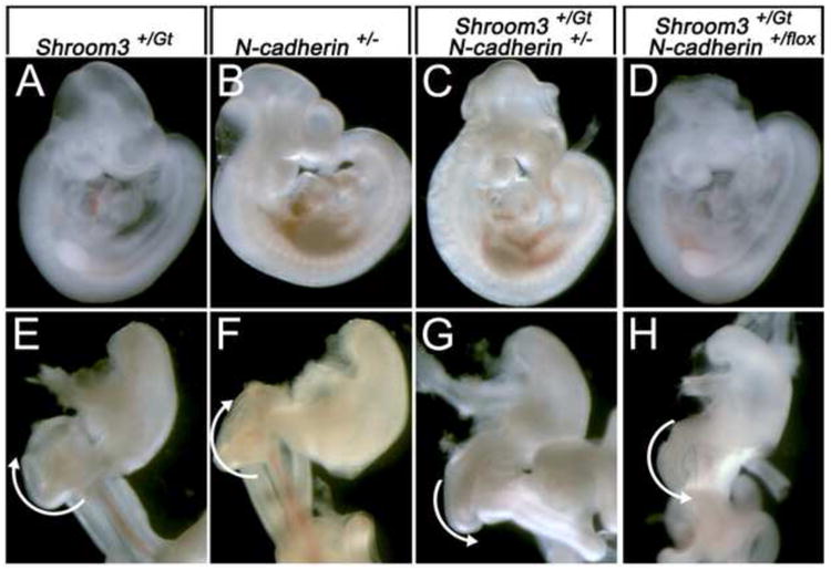
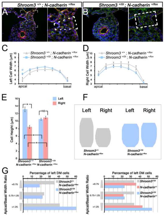
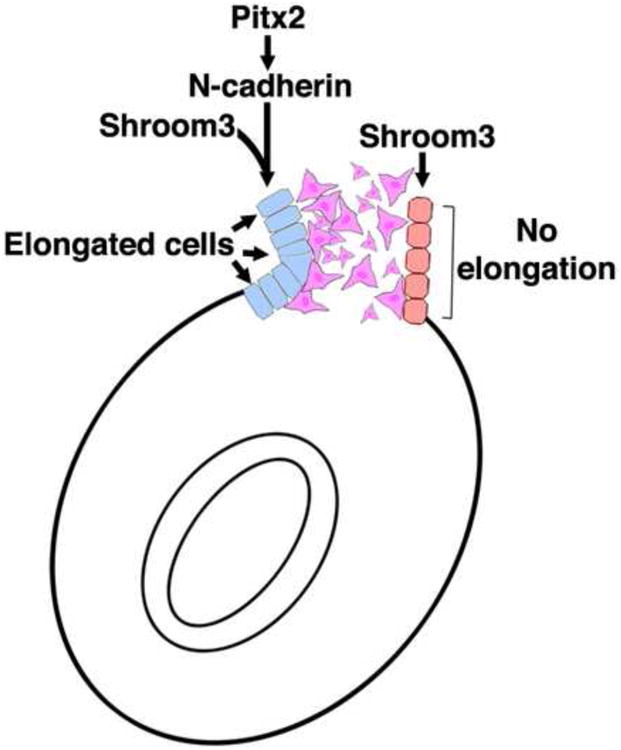
The cytoskeletal protein Shroom3 is a potent inducer of epithelial cell shape change and is required for lens and neural plate morphogenesis. Analysis of gut morphogenesis in Shroom3 deficient mouse embryos revealed that the direction of gut rotation is also disrupted. It was recently established that Pitx2-dependent, asymmetrical cellular behaviors in the dorsal mesentery (DM) of the early mid-gut, a structure connecting the gut-tube to the rest of the embryo, contribute to the direction of gut rotation in chicken embryos by influencing the direction of the dorsal mesenteric tilt. Asymmetric cell shapes in the DM epithelium are hypothesized to contribute to the tilt, however, it is unclear what lies downstream of Pitx2 to alter epithelial cell shape. The cells of the left DM epithelium in either Pitx2 or Shroom3 deficient embryos are shorter and wider than those in control embryos and resemble the shape of those on the right, demonstrating that like Pitx2, Shroom3 is required for cell shape asymmetry and the leftward DM tilt. Because N-cadherin expression is specific to the left side and is Pitx2 dependent, we determined whether Shroom3 and N-cadherin function together to regulate cell shape in the left DM epithelium. Analysis of mouse embryos lacking one allele of both Shroom3 and N-cadherin revealed that they possess shorter and wider left epithelial DM cells when compared with Shroom3 or N-cadherin heterozygous embryos. This indicates a genetic interaction. Together these data provide evidence that Shroom3 and N-cadherin function cooperatively downstream of Pitx2 to directly regulate cell shape changes necessary for early gut tube morphogenesis.


Figures

Figure 1
Figure 1. Shroom3 and Pitx2 deficient mice have similar gut coiling defects
(A-J) Ventral view of digestive tracts from embryos of the indicated age and genotype were dissected from the liver and pancreas in order to visualize the direction of gut coiling (white arrows). The outlines of the Stomach (St), Duodenum (Du), and proximal intestines are diagrammed at E15.5 in G and H to illustrate directional coiling.
Figure 2
Figure 2. Shroom3 and Pitx2 are required for cell shape asymmetries during gut morphogenesis
(A-F) E10.75 embryos of the indicated genotype were transversely cyrosectioned caudally, approximately halfway between the fore- and hind-limbs (A,C,E), or rostrally near the duodenum (B,D,F) and immunofluorescently labeled with F-actin (green) and/or β-catenin (red). The area indicated in B, D, and F is magnified and displayed in the right-most panels. The dashed white outlines are traces of β-catenin signal demarcating individual cell borders. (G-I) Traces of the left and right dorsal mesentery epithelial cell outlines were measured and the average widths along the apical-basal axis (G,H) and height (I) were calculated. Asterisks represent statistical significance compared with the control (wt) averages (p<0.05). (J) The average widths and height of left and right dorsal mesentery epithelial cells from the indicated genotypes was quantified and used to generate diagrams representing the average cell shape. (K) Representative image of wild-type left dorsal mesentery epithelium containing apically constricted cells (arrows). The diagram on the right outlines the shape of the cells pictured with asterisks marking apically constricted cells. (L) The number of left DM epithelial cells from embryos of the indicated genotypes, defined by those with an apical/basal width ratio of <0.75, 0.75-1.25, or >1.25 indicating apically constricted, columnar, or basally constricted shapes, respectively, were counted and the total percentage of each type of cell was determined.
Figure 3
Figure 3. F-actin and Myosin II are mislocalized in the dorsal mesentery of Shroom3 deficient embryos
(A-D) E10.75 wild-type or Shroom3Gt/Gt embryos were cryosectioned just caudal to the duodenum and the sections were F-actin and β-catenin immunofluorescently labeled. The boxed region indicates the region magnified in the adjacent panel. The arrowheads indicate regions of intense labeling. (E-F) The average apical/basal F-actin and Myosin II intensity ratios from wild-type and Shroom3Gt/Gt embryos were calculated from the left and right dorsal mesentery epithelium. Asterisks and brackets indicate means that are significantly different (p<0.05). NS=not significant
Figure 4
Figure 4. Pitx2 regulates N-cadherin but not Shroom3 in the left dorsal mesentery epithelium
(A) X-gal staining of the dorsal mesentery from E10.75 Shroom3+/Gt embryos. (B) Shroom3+/Gt mice were crossed with the Z/EG line, which reports cre-recombinase activity via GFP expression. E10.75 embryos from this cross were visualized for GFP to observe areas of the dorsal mesentery expressing Cre under control of the Shroom3 locus. (C-H) E10.75 embryos of the indicated genotyped were cryosectioned and immunofluorescently labeled with the indicated antibody. The bracketed region indicates the area magnified in the adjacent panel. The arrows indicate regions of strong labeling intensity and the dotted line indicates the border of strong expression.
Figure 5
Figure 5. N-cadherin and Shroom3 cooperatively function during neural tube and gut morphogenesis
(A-D) Whole embryos with the indicated genotypes are indicated. Note the presence of exencephaly in embryos in panels C and D. (E-H) Ventral view of digestive tracts dissected from E14.5 embryos of the indicated genotype dissected from the liver and pancreas. The white arrows indicate the direction of gut coiling.
Figure 6
Figure 6. N-cadherin and Shroom3 cooperatively regulate cell shape in the dorsal mesentery
(A-B) Representative cryosections from E10.75 embryos with the indicated genotype were immunofluorescently labeled with β-catenin (red), F-actin (green), and Hoechst (blue). The hatched lines indicate the general shape of the dorsal mesentery; note the trapezoidal shape in the control (A) and the rectangular shape in the mutant embryo (B). (C-E) Individual cell shapes of E10.75 left and right dorsal mesentery epithelia of the indicated genotypes were outlined and their average height and width were quantified. Asterisks and brackets represent a significant difference compared with the control (wt) averages (p<0.05). NS=not significant (F) The average cell shape of dorsal mesentery epithelial cells from the indicated genotype was generated utilizing the averages quantified in C-E and depicted. (G) The number of left DM epithelial cells from embryos of the indicated genotypes, defined by those with an apical/basal width ratio of <0.75, 0.75-1.25, or >1.25 indicating apically constricted, columnar, or basally constricted shapes, respectively, were counted and the total percentage of each type of cell was determined.
Figure 7
Figure 7. Shroom3 and a Pitx2-N-cadherin pathway function cooperatively to generate asymmetric cell shape changes during gut morphogenesis
The model illustrates the proposed mechanism where Shroom3 regulates cell shape specifically in the left dorsal mesentery downstream of Pitx2. The Pitx2 regulation of Shroom3 activity is achieved by directing asymmetric, leftward N-cadherin expression. The elongation and apical constriction of left epithelial dorsal mesentery cells together with asymmetrical mesenchymal packing causes leftward tilting biasing the direction of gut coiling.

Similar articles

Cited by

References

    1. Ai D, Liu W, Ma L, Dong F, Lu MF, Wang D, Verzi MP, Cai C, Gage PJ, Evans S, et al. Pitx2 regulates cardiac left-right asymmetry by patterning second cardiac lineage-derived myocardium. Dev Biol. 2006;296(2):437–49. - PMC - PubMed
    1. Campione M, Steinbeisser H, Schweickert A, Deissler K, van Bebber F, Lowe LA, Nowotschin S, Viebahn C, Haffter P, Kuehn MR, et al. The homeobox gene Pitx2: mediator of asymmetric left-right signaling in vertebrate heart and gut looping. Development. 1999;126(6):1225–34. - PubMed
    1. Chung MI, Nascone-Yoder NM, Grover SA, Drysdale TA, Wallingford JB. Direct activation of Shroom3 transcription by Pitx proteins drives epithelial morphogenesis in the developing gut. Development. 2010;137(8):1339–49. - PMC - PubMed
    1. Davis NM, Kurpios NA, Sun X, Gros J, Martin JF, Tabin CJ. The chirality of gut rotation derives from left-right asymmetric changes in the architecture of the dorsal mesentery. Dev Cell. 2008;15(1):134–45. - PMC - PubMed
    1. Gage PJ, Suh H, Camper SA. Dosage requirement of Pitx2 for development of multiple organs. Development. 1999;126(20):4643–51. - PubMed

Publication types

MeSH terms