. 2011 Nov;23(11):1794-805.
doi: 10.1016/j.cellsig.2011.06.014. Epub 2011 Jun 25.

Prolactin-stimulated activation of ERK1/2 mitogen-activated protein kinases is controlled by PI3-kinase/Rac/PAK signaling pathway in breast cancer cells

Affiliations

Prolactin-stimulated activation of ERK1/2 mitogen-activated protein kinases is controlled by PI3-kinase/Rac/PAK signaling pathway in breast cancer cells

Edita Aksamitiene et al. Cell Signal. 2011 Nov.

Abstract

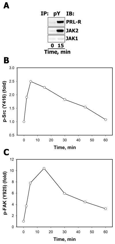
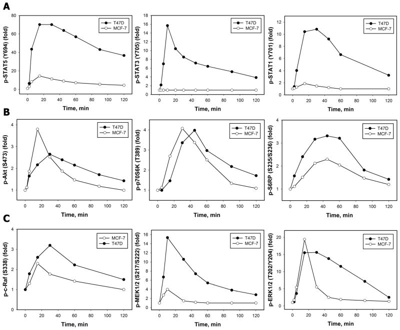
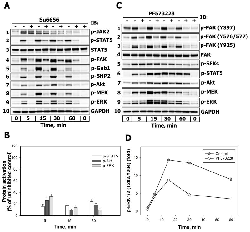
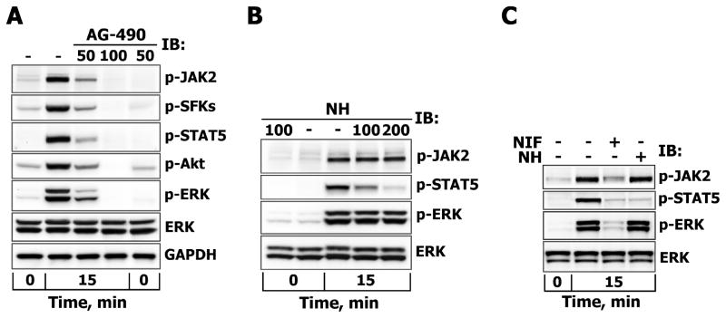
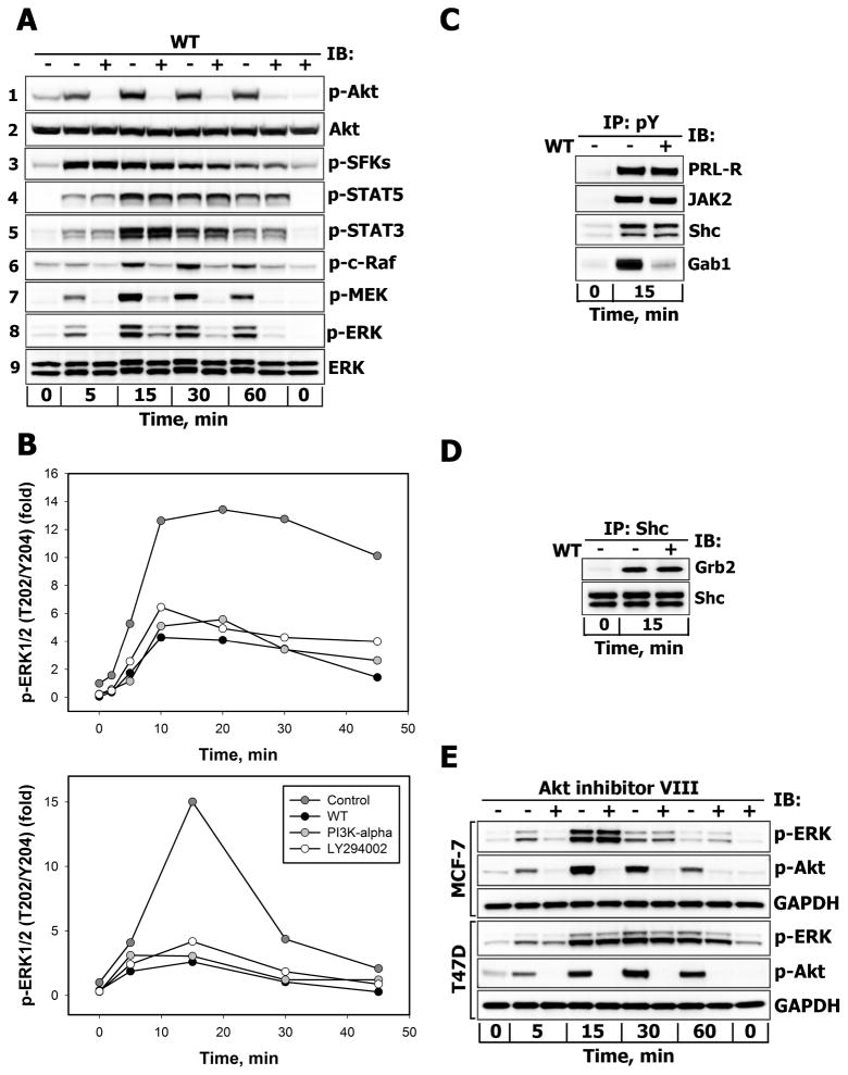
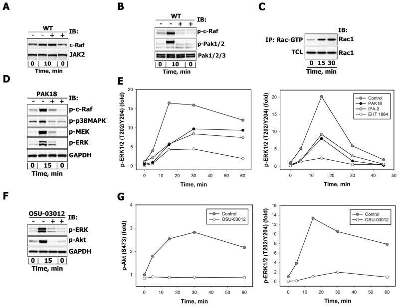
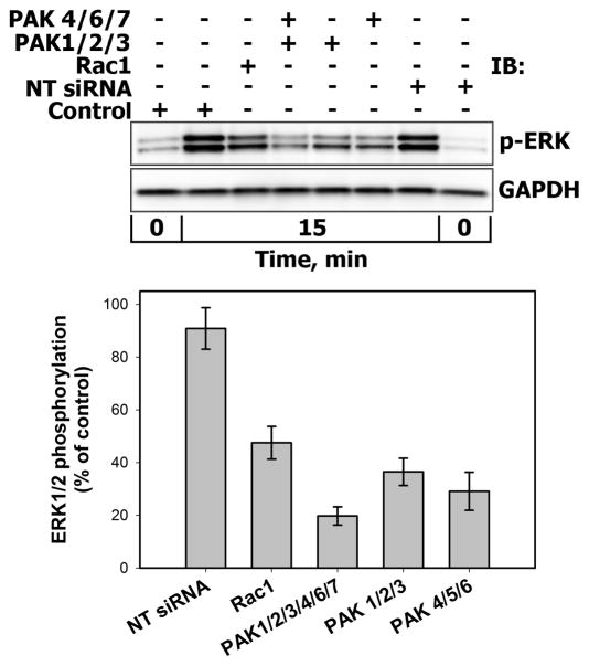
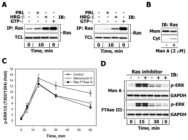
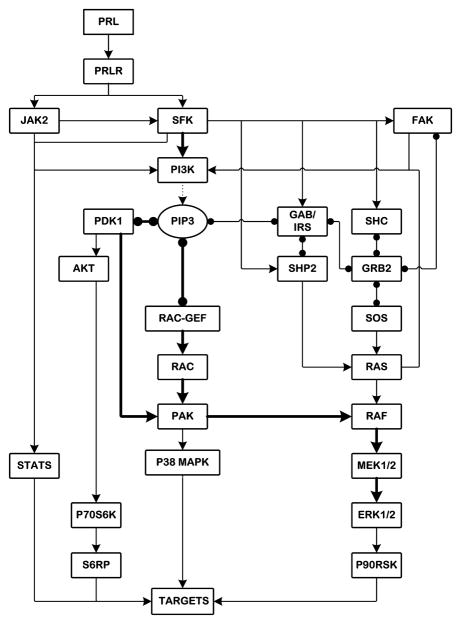
There is strong evidence that deregulation of prolactin (PRL) signaling contributes to pathogenesis and chemoresistance of breast cancer. Therefore, understanding cross-talk between distinct signal transduction pathways triggered by activation of the prolactin receptor (PRL-R), is essential for elucidating the pathogenesis of metastatic breast cancer. In this study, we applied a sequential inhibitory analysis of various signaling intermediates to examine the hierarchy of protein interactions within the PRL signaling network and to evaluate the relative contributions of multiple signaling branches downstream of PRL-R to the activation of the extracellular signal-regulated kinases ERK1 and ERK2 in T47D and MCF-7 human breast cancer cells. Quantitative measurements of the phosphorylation/activation patterns of proteins showed that PRL simultaneously activated Src family kinases (SFKs) and the JAK/STAT, phosphoinositide-3 (PI3)-kinase/Akt and MAPK signaling pathways. The specific blockade or siRNA-mediated suppression of SFK/FAK, JAK2/STAT5, PI3-kinase/PDK1/Akt, Rac/PAK or Ras regulatory circuits revealed that (1) the PI3-kinase/Akt pathway is required for activation of the MAPK/ERK signaling cascade upon PRL stimulation; (2) PI3-kinase-mediated activation of the c-Raf-MEK1/2-ERK1/2 cascade occurs independent of signaling dowstream of STATs, Akt and PKC, but requires JAK2, SFKs and FAK activities; (3) activated PRL-R mainly utilizes the PI3-kinase-dependent Rac/PAK pathway rather than the canonical Shc/Grb2/SOS/Ras route to initiate and sustain ERK1/2 signaling. By interconnecting diverse signaling pathways PLR may enhance proliferation, survival, migration and invasiveness of breast cancer cells.


Figures

Figure 1
Figure 1
A. PRL induced activation of PRL-R and JAK family tyrosine kinases Serum-starved T47D cells were either left unstimulated or were stimulated with 10 nM PRL for 15 min. Tyrosine phosphorylated proteins were immunoprecipitated (IP) from total cell lysates (TCL) using the monoclonal agarose-conjugated anti-phosphotyrosine antibody (Ab), pY-20. Resolved proteins were transferred onto nitrocellulose membrane, which was immunoblotted (IB) with Abs against the proteins, indicated on the right. B–C. PRL activates Src family tyrosine kinases and their target FAK. Total and phosphorylated forms of Src family protein kinases (B) and FAK (C) of unstimulated and PRL-stimulated (10 nM, for the indicated time intervals) T47D cells were detected by IB of the TCL with anti-c-Src, anti-phospho-Src (Tyr416), anti-FAK and anti-phospho-FAK (Tyr925) Abs, respectively. The signal intensities of phosphorylated protein were normalized by signal intensities of the total (phosphorylated and non-phosphorylated) protein at each time point and then expressed as fold changes over basal levels (in unstimulated cells). Densitometric quantitation of representative blots is shown (n=3).
Figure 2
Figure 2. Time-courses of PRL-induced activation of STAT (A), PI3-kinase (B) and MAPK (C) signaling pathways in breast cancer cells
Both total and phosphorylated forms of STAT5 (Tyr694) (A, left panel), STAT3 (Tyr705) (A, middle panel), STAT1 (Tyr701) (A, right panel), Akt (Ser473) (B, left panel), p70S6K (Thr389) (B, middle panel), S6RP (Ser235/Ser236) (B, right panel), c-Raf (Ser338) (C, left panel), MEK1/2 (Ser217) (C, middle panel) and ERK1/2 (Thr202/Tyr204) (C, right panel) proteins were detected by IB with Abs against respective antigens in TCL of unstimulated and PRL-stimulated (10 nM, for the indicated time intervals) T47D (black circles) or MCF-7 (white circles) cells. The ratio of phospho-protein:total protein at each time point was expressed as fold changes over basal levels. Quantitation of representative blots is shown (n=3).
Figure 3
Figure 3
A–B. The effects of SFK inhibition on the activation of downstream effectors of PRL-R Serum-starved T47D (A) or MCF-7 (B) cells were either left untreated (−) or were treated (+) with Su6656 (10 μM, 30 min) before stimulation with 10 nM PRL for the indicated time intervals. Phosphorylated forms of JAK2 (Tyr Tyr1007/1008), STAT5 (Tyr694), FAK (Tyr925), Gab1 (Tyr627), SHP2 (Tyr542), Akt (Ser473), MEK1/2 (Ser217/Ser221) and ERK1/2 (Thr202/Tyr204) proteins were detected by IB of the TCL with Abs against respective antigens. GAPDH levels were detected with anti-GAPDH Abs and used as protein loading control. Representative blots are shown (n=3). B. Phosphorylated STAT5 (white bars), Akt (dark grey bars) and ERK1/2 (light grey bars) in TCL of Su6656-treated cells were quantified by densitometry and are expressed as percent of control of the corresponding phospho-protein measured in TCL of PRL-stimulated cells incubated without Su6656. Data represent the mean ± SD of three independent experiments. C–D. The effects of FAK inhibition on the activation of downstream effectors of PRL-R. Serum-starved T47D cells were either left untreated (−) or were treated (+) with PF573228 (0.5 μM, 2 h) before stimulation with 10 nM PRL for the indicated time intervals. Phosphorylated forms of FAK (Tyr397), FAK (Tyr576/577), FAK (Tyr925), SFKs (Tyr416), STAT5 (Tyr694), Akt (Ser473), MEK1/2 (Ser217/Ser221) and ERK1/2 (Thr202/Tyr204) proteins were detected by IB of the TCL with Abs against respective antigens. GAPDH levels were used as protein loading control. Representative blots are shown (n=3). D. The ratio of phospho-ERK1/2:total ERK1/2 at each time point was expressed as fold changes over basal levels.
Figure 4
Figure 4. The effects of JAK2 and STAT5 inhibition on PRL-induced activation of ERK1/2
Serum-starved T47D cells were either left untreated (−) or were treated (+) with AG-490 (50 or 100 μM) for 3 hours (A) or with STAT5 inhibitor (NH, 100 or 200 μM) for 1 hour (B), while MCF-7 cells were treated with NH (200 μM) or nifuroxazide (NIF, 100 μM) for 1 hour (C) before stimulation with 10 nM PRL for 15 min. Phosphorylated forms of JAK2 (Tyr1007/1008), SFKs (Tyr416), Akt (Ser473), STAT5 (Tyr694) and ERK1/2 (Thr202/Tyr204) proteins were detected by IB of the TCL with Abs against respective antigens. Total ERK1/2 protein levels were detected with anti-ERK1/2 Abs and were used as protein loading control. Representative blots are shown.
Figure 5
Figure 5
A. Effects of PI3-kinase inhibition on the activation of downstream effectors of PRL-R Serum-starved T47D cells were either left untreated (−) or were treated (+) with wortmannin (WT, 200 nM, 30 min) before stimulation with 10 nM PRL for the indicated time intervals. Phosphorylated forms of Akt (Ser473), SFKs (Tyr416), STAT5 (Tyr694), STAT3 (Tyr705), c-Raf (Ser338), MEK1/2 (Ser217/Ser221) and ERK1/2 (Thr202/Tyr204) proteins were detected by IB of the TCL with Abs against respective antigens. Total Akt and ERK1/2 protein levels were detected with anti-Akt1 or anti-ERK1/2 Abs, respectively, and served as protein loading controls. Representative blots are shown (n=3). B. Effects of structurally different inhibitors of PI3-kinase on PRL-induced activation of ERK1/2. Serum-starved T47D (upper panel) and MCF-7 (lower panel) cells were either left untreated (control, dark grey circles) or were preincubated with WT (200 nM, black circles), PI3K-α inhibitor 2 (2 μM, grey circles) or LY 294002 (25 μM, white circles) for 30 min before stimulation with 10 nM PRL for the indicated time intervals. Both total and phosphorylated forms of ERK1/2 were detected by IB of the TCL with anti-ERK1/2 or anti-phospho-ERK1/2 (Thr202/Tyr204) Abs, respectively. The ratio of phospho-ERK1/2:total ERK1/2 at each time point is expressed as fold changes over basal levels. Quantitation of represenatative blots is shown (n=3). C. Effects of PI3-kinase inhibition on tyrosine phosphorylation levels of PRL-R, JAK2, Shc and Gab proteins. Tyrosine phosphorylated proteins were immunoprecipitated (IP) from TCL of unstimulated or PRL-stimulated (10 nM, 15 min) and WT-treated (+) (200 nM, 30 min) or untreated (−) T47D cells and probed for proteins indicated on the right. D. Effects of PI3-kinase inhibition on Shc association with Grb2. Shc proteins were immunoprecipitated (IP) from TCL of unstimulated or PRL-stimulated (10 nM, 15 min) and WT-treated (+) (200 nM, 30 min) or untreated (−) T47D cells and probed with anti-Grb2 or anti-Shc Abs. E. Effects of Akt inhibition on PRL-induced activation of ERK1/2. Serum-starved MCF-7 or T47D cells were either left untreated (−) or were treated (+) with Akt1/2/3 inhibitor (Akt-VIII, 10 μM, 30 min) before stimulation with 10 nM PRL for the indicated time intervals. Phosphorylated forms of Akt (Ser473) and ERK1/2 (Thr202/Tyr204) proteins were detected by IB of the TCL with Abs against respective antigens. GAPDH levels served as protein loading control. Representative blots are shown (n=3).
Figure 6
Figure 6
A. Effects of PI3-kinase inhibition on PRL-induced recruitment of c-Raf Serum-starved T47D cells were either left untreated (−) or were treated (+) with WT (200 nM, 30min) before stimulation with 10 nM PRL for 10 min. c-Raf proteins were detected by IB of the particulate/membrane fraction, which was isolated as described in “Materials and Methods”. Simultaneous detection of JAK2 in the same samples served as loading control. B. Effects of PI3K inhibition on PRL-induced c-Raf and PAK activation. Serum-starved T47D cells were either left untreated (−) or were treated (+) with wortmannin (WT, 200 nM, 30 min) before stimulation with PRL (10 nM, 10 min). Phosphorylated c-Raf (Ser338), PAK1/2 (Thr423/Thr402) and total PAK1/2/3 protein levels were detected by IB with Abs against respective antigens. C. PRL stimulation of Rac1 activation. After MCF-7 cell stimulation with 10 nM PRL, Rac-GTP levels were measured at indicated time points by affinity precipitation with p21-Rac1 binding domain of human PAK1 bound to glutathione agarose beads as described in “Materials and Methods” and detected with anti-Rac1Abs, which were also used to determine total Rac1 protein levels in corresponding TCL. D. Effects of PAK-18 on PRL-induced activation of MAPK cascades. Serum-starved T47D cells were either left untreated (−) or were treated (+) with PAK18 peptide (10 μM, 60 min) before stimulation with 10 nM PRL for 15 min. Phosphorylated forms of c-Raf (Ser338), p38MAPK (Thr180/Tyr182), MEK1/2 (Ser217/221) and ERK1/2 (Thr202/Tyr204) proteins were detected by IB of the TCL with Abs against respective antigens. GAPDH levels served as protein loading control. Representative blots are shown (n=3). E. Effects of Rac/PAK inhibition on PRL-induced activation of ERK1/2. Serum-starved T47D (left panel) or MCF-7 (right panel) cells were either left untreated (control, dark grey circles) or were treated (+) with EHT 1864 (10 μM, 60 min, white circles), IPA-3 (10 μM, 30 min, grey circles) or PAK18 peptide (10 μM, 60 min, black circles) before stimulation with 10 nM PRL for the indicated time intervals. Both total and phosphorylated forms of ERK1/2 (Thr202/Tyr204) were detected by IB of the TCL with Abs against respective antigens. The ratio of phospho-ERK1/2:total ERK1/2 at each time point was expressed as fold changes over basal levels. Quantitation of representative blots is shown (n=3). F–G. Effects of concurrent inhibition of PDK1 and PAK on PRL-induced activation of Akt and ERK1/2. Serum-starved MCF-7 (F) or T47D (G) cells were either left untreated (−) or were treated (+) with OSU-03012 (25 μM, 30 min) before stimulation with 10 nM PRL for indicated periods of time. Phosphorylated forms of Akt (Ser473) and ERK1/2 (Thr202/Tyr204) proteins were detected by IB of the TCL with Abs against respective antigens. GAPDH levels served as protein loading control. Representative blots are shown (n=3). (F). The ratio of phospho-protein:total protein at each time point was expressed as fold changes over basal levels (G).
Figure 7
Figure 7. Effects of siRNA-mediated suppression of Rac1 and PAK family members on PRL-induced activation of ERK1/2
MCF-7 cells were either left untrasfected (Control) or were transiently transfected in dublicates with non-targeting (NT) negative control siRNA, siRNA duplexes specific for Rac1, group I PAKs (PAK1/2/3), group II PAKs (PAK4/6/7) or group I/II PAKs for 72 hours. Cells were then stimulated with 10 nM PRL for 15 min. Phosphorylated forms of ERK1/2 (Thr202/Tyr204) were detected by IB of the TCL. GAPDH levels served as protein loading control. Representative blots are shown in the upper panel. The lower panel shows a graph with the calculated averages ±SD of remaining ERK1/2 phosphorylation (normalized to GAPDH) from two independent experiments.
Figure 8
Figure 8
A. Comparison of Ras activation in PRL and HRG-β-treated breast cancer cells Serum-starved T47D (left panel) or MCF-7 (right panel) were either left unstimulated or were stimulated with 10 nM PRL or HRG-β for 10 min. GTP-gamma-S loading of Ras in non-stimulated cells was used as a positive control for Ras activation. Affinity precipitation of activated Ras was carried out as described in “Materials and Methods”. Ras was detected by IB of the TCL and corresponding immunoprecipitates (IP) with monoclonal anti-Ras Abs. B. Effects of the farnesyl transferase inhibitor manumycin A on membrane accumulation of Ras. T47D cells were either left untreated (−) or were treated with manumycin A (2 μM) for 7 hours in serum-free medium. Ras was detected by IB of cytosolic (Cyt) and particulate/membrane (Mem) fractions with monoclonal anti-Ras Abs. C–D. Effects of farnesyl transferase inhibitors on PRL-induced ERK1/2 activation. C. Serum-starved T47D cells were either left untreated (control, grey circles) or were treated with RasFTase III (2 μM, black circles) or manumycin A (2 μM, white circles) for 7 hours before stimulation with 10 nM PRL for indicated time intervals. Both total and phosphorylated forms of ERK1/2 (Thr202/Tyr204) were detected by IB with Abs against respective antigens. The ratio of phospho-ERK1/2:total ERK1/2 at each time point was expressed as fold changes over basal levels. Quantitation of representative blots is shown (n=3). D. Phosphorylated ERK1/2 and total GAPDH proteins were detected by IB the TCL of similarly treated MCF-7 cells. Representative blots are shown (n=3).
Figure 9
Figure 9. Suggested architecture of PRL-R signaling network with potential routes of MAPK activation in breast cancer cells
Solid lines with arrows show the activation or phosphorylation of proteins. Dotted lines indicate the formation of phosphatidylinositol-trisphosphate (PIP3) by PI3-kinase. Solid lines with circular ends represent protein-protein and protein-lipid interactions. Thick solid lines show prevailing routes of ERK1/2 activation.

References

    1. Binart N, Ormandy CJ, Kelly PA. Adv Exp Med Biol. 2000;480:85–92. - PubMed
    1. Fresno Vara JA, Caceres MA, Silva A, Martin-Perez J. Mol Biol Cell. 2001;12(7):2171–2183. - PMC - PubMed
    1. Clevenger CV, Furth PA, Hankinson SE, Schuler LA. Endocr Rev. 2003;24(1):1–27. - PMC - PubMed
    1. Peirce SK, Chen WY. J Endocrinol. 2001;171(1):R1–4. - PubMed
    1. Bernichtein S, Touraine P, Goffin V. J Endocrinol. 2010;206(1):1–11. - PubMed

Publication types

MeSH terms